4RKG

Structure of the MSL2 CXC domain bound with a non-specific (GC)6 DNA


Experimental Data Snapshot

  • Method: X-RAY DIFFRACTION
  • Resolution: 2.50 Å
  • R-Value Free: 0.315 
  • R-Value Work: 0.227 
  • R-Value Observed: 0.231 

wwPDB Validation   3D Report Full Report


This is version 1.0 of the entry. See complete history


Literature

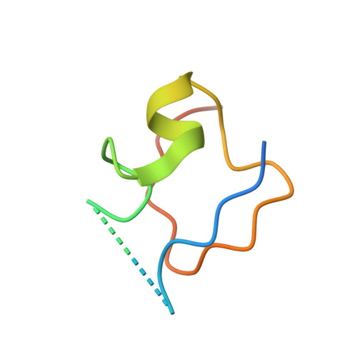
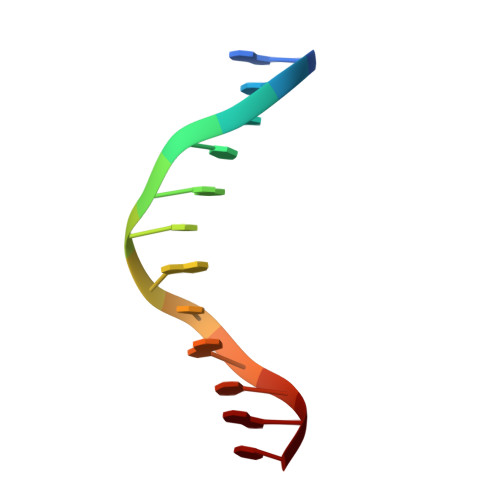
Structural basis of X chromosome DNA recognition by the MSL2 CXC domain during Drosophila dosage compensation.

Zheng, S.Villa, R.Wang, J.Feng, Y.Wang, J.Becker, P.B.Ye, K.

(2014) Genes Dev 28: 2652-2662

  • DOI: https://doi.org/10.1101/gad.250936.114
  • Primary Citation of Related Structures:  
    4RKG, 4RKH

  • PubMed Abstract: 

    The male-specific lethal dosage compensation complex (MSL-DCC) selectively assembles on the X chromosome in Drosophila males and activates gene transcription by twofold through histone acetylation. An MSL recognition element (MRE) sequence motif nucleates the initial MSL association, but how it is recognized remains unknown. Here, we identified the CXC domain of MSL2 specifically recognizing the MRE motif and determined its crystal structure bound to specific and nonspecific DNAs. The CXC domain primarily contacts one strand of DNA duplex and employs a single arginine to directly read out dinucleotide sequences from the minor groove. The arginine is flexible when bound to nonspecific sequences. The core region of the MRE motif harbors two binding sites on opposite strands that can cooperatively recruit a CXC dimer. Specific DNA-binding mutants of MSL2 are impaired in MRE binding and X chromosome localization in vivo. Our results reveal multiple dynamic DNA-binding modes of the CXC domain that target the MSL-DCC to X chromosomes.


  • Organizational Affiliation

    Department of Biochemistry and Molecular Biology, College of Life Sciences, Beijing Normal University, Beijing 100875, China; National Institute of Biological Sciences, Beijing 102206, China;


Macromolecules

Find similar proteins by:  (by identity cutoff)  |  3D Structure
Entity ID: 1
MoleculeChains Sequence LengthOrganismDetailsImage
E3 ubiquitin-protein ligase msl-2
A, B
52Drosophila melanogasterMutation(s): 1 
Gene Names: CG3241msl-2MSL2
EC: 6.3.2
UniProt
Find proteins for P50534 (Drosophila melanogaster)
Explore P50534 
Go to UniProtKB:  P50534
Entity Groups  
Sequence Clusters30% Identity50% Identity70% Identity90% Identity95% Identity100% Identity
UniProt GroupP50534
Sequence Annotations
Expand
  • Reference Sequence

Find similar nucleic acids by:  Sequence   |   3D Structure  

Entity ID: 2
MoleculeChains LengthOrganismImage
DNA (5'-D(*GP*CP*GP*CP*GP*CP*GP*CP*GP*CP*GP*C)-3')C [auth H],
D [auth I]
12N/A
Sequence Annotations
Expand
  • Reference Sequence
Experimental Data & Validation

Experimental Data

  • Method: X-RAY DIFFRACTION
  • Resolution: 2.50 Å
  • R-Value Free: 0.315 
  • R-Value Work: 0.227 
  • R-Value Observed: 0.231 
  • Space Group: P 21 21 21
Unit Cell:
Length ( Å )Angle ( ˚ )
a = 40.705α = 90
b = 75.251β = 90
c = 77.436γ = 90
Software Package:
Software NamePurpose
ADSCdata collection
PHASERphasing
PHENIXrefinement
DENZOdata reduction
SCALEPACKdata scaling

Structure Validation

View Full Validation Report


Entry History 

Deposition Data

Revision History  (Full details and data files)

  • Version 1.0: 2015-01-21
    Type: Initial release