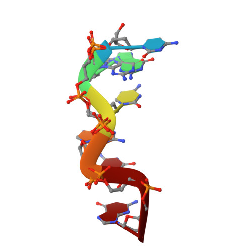
High-resolution crystal structure of Z-DNA in complex with Cr(3+) cations.
Drozdzal, P., Gilski, M., Kierzek, R., Lomozik, L., Jaskolski, M.(2015) J Biol Inorg Chem 20: 595-602
- PubMed: 25687556
- DOI: https://doi.org/10.1007/s00775-015-1247-5
- Primary Citation of Related Structures:
4R15 - PubMed Abstract:
This work is part of our project aimed at characterizing metal-binding properties of left-handed Z-DNA helices. The three Cr(3+) cations found in the asymmetric unit of the d(CGCGCG)2-Cr(3+) crystal structure do not form direct coordination bonds with atoms of the Z-DNA molecule. Instead, the hydrated Cr(3+) ions are engaged in outer-sphere interactions with phosphate groups and O6 and N7 guanine atoms of the DNA. The Cr(3+)(1) and Cr(3+)(2) ions have disordered coordination spheres occupied by six water molecules each. These partial-occupancy chromium cations are 2.354(15) Å apart and are bridged by three water molecules from their hydration spheres. The Cr(3+)(3) cation has distorted square pyramidal geometry. In addition to the high degree of disorder of the DNA backbone, alternate conformations are also observed for the deoxyribose and base moieties of the G2 nucleotide. Our work illuminates the question of conformational flexibility of Z-DNA and its interaction mode with transition-metal cations.
Organizational Affiliation:
Faculty of Chemistry, A. Mickiewicz University, Umultowska 89b, 61-614, Poznan, Poland.


