4QMA

Crystal Structure of a Putative Cysteine Dioxygnase From Ralstonia eutropha: An Alternative Modeling of 2GM6 from JCSG Target 361076


Experimental Data Snapshot

  • Method: X-RAY DIFFRACTION
  • Resolution: 1.65 Å
  • R-Value Free: 0.200 
  • R-Value Work: 0.174 
  • R-Value Observed: 0.175 

wwPDB Validation   3D Report Full Report


This is version 1.5 of the entry. See complete history


Literature

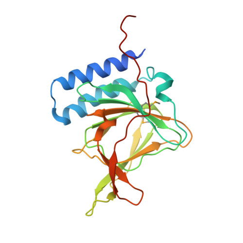
Structures of Arg- and Gln-type bacterial cysteine dioxygenase homologs.

Driggers, C.M.Hartman, S.J.Karplus, P.A.

(2015) Protein Sci 24: 154-161

  • DOI: https://doi.org/10.1002/pro.2587
  • Primary Citation of Related Structures:  
    4QM8, 4QM9, 4QMA

  • PubMed Abstract: 

    In some bacteria, cysteine is converted to cysteine sulfinic acid by cysteine dioxygenases (CDO) that are only ∼15-30% identical in sequence to mammalian CDOs. Among bacterial proteins having this range of sequence similarity to mammalian CDO are some that conserve an active site Arg residue ("Arg-type" enzymes) and some having a Gln substituted for this Arg ("Gln-type" enzymes). Here, we describe a structure from each of these enzyme types by analyzing structures originally solved by structural genomics groups but not published: a Bacillus subtilis "Arg-type" enzyme that has cysteine dioxygenase activity (BsCDO), and a Ralstonia eutropha "Gln-type" CDO homolog of uncharacterized activity (ReCDOhom). The BsCDO active site is well conserved with mammalian CDO, and a cysteine complex captured in the active site confirms that the cysteine binding mode is also similar. The ReCDOhom structure reveals a new active site Arg residue that is hydrogen bonding to an iron-bound diatomic molecule we have interpreted as dioxygen. Notably, the Arg position is not compatible with the mode of Cys binding seen in both rat CDO and BsCDO. As sequence alignments show that this newly discovered active site Arg is well conserved among "Gln-type" CDO enzymes, we conclude that the "Gln-type" CDO homologs are not authentic CDOs but will have substrate specificity more similar to 3-mercaptopropionate dioxygenases.


  • Organizational Affiliation

    Department of Biochemistry and Biophysics, 2011 Ag & Life Sciences Bldg, Oregon State University, Corvallis, Oregon, 97331.


Macromolecules
Find similar proteins by:  (by identity cutoff)  |  3D Structure
Entity ID: 1
MoleculeChains Sequence LengthOrganismDetailsImage
Cysteine dioxygenase type I208Cupriavidus pinatubonensis JMP134Mutation(s): 0 
Gene Names: Reut_B5045YP_299237.1
UniProt
Find proteins for Q46R41 (Cupriavidus pinatubonensis (strain JMP 134 / LMG 1197))
Explore Q46R41 
Go to UniProtKB:  Q46R41
Entity Groups  
Sequence Clusters30% Identity50% Identity70% Identity90% Identity95% Identity100% Identity
UniProt GroupQ46R41
Sequence Annotations
Expand
  • Reference Sequence
Small Molecules
Ligands 4 Unique
IDChains Name / Formula / InChI Key2D Diagram3D Interactions
SO4
Query on SO4

Download Ideal Coordinates CCD File 
D [auth A],
E [auth A]
SULFATE ION
O4 S
QAOWNCQODCNURD-UHFFFAOYSA-L
EDO
Query on EDO

Download Ideal Coordinates CCD File 
F [auth A]
G [auth A]
H [auth A]
I [auth A]
J [auth A]
F [auth A],
G [auth A],
H [auth A],
I [auth A],
J [auth A],
K [auth A],
L [auth A],
M [auth A],
N [auth A]
1,2-ETHANEDIOL
C2 H6 O2
LYCAIKOWRPUZTN-UHFFFAOYSA-N
FE
Query on FE

Download Ideal Coordinates CCD File 
B [auth A]FE (III) ION
Fe
VTLYFUHAOXGGBS-UHFFFAOYSA-N
OXY
Query on OXY

Download Ideal Coordinates CCD File 
C [auth A]OXYGEN MOLECULE
O2
MYMOFIZGZYHOMD-UHFFFAOYSA-N
Modified Residues  1 Unique
IDChains TypeFormula2D DiagramParent
MSE
Query on MSE
A
L-PEPTIDE LINKINGC5 H11 N O2 SeMET
Experimental Data & Validation

Experimental Data

  • Method: X-RAY DIFFRACTION
  • Resolution: 1.65 Å
  • R-Value Free: 0.200 
  • R-Value Work: 0.174 
  • R-Value Observed: 0.175 
  • Space Group: P 41 21 2
Unit Cell:
Length ( Å )Angle ( ˚ )
a = 57.007α = 90
b = 57.007β = 90
c = 216.683γ = 90
Software Package:
Software NamePurpose
ADSCdata collection
PHENIXmodel building
PHENIXrefinement
MOSFLMdata reduction
SCALAdata scaling
PHENIXphasing

Structure Validation

View Full Validation Report


Entry History 

Deposition Data

Revision History  (Full details and data files)

  • Version 1.0: 2014-11-26
    Type: Initial release
  • Version 1.1: 2014-12-31
    Changes: Database references
  • Version 1.2: 2015-01-14
    Changes: Database references
  • Version 1.3: 2015-04-29
    Changes: Structure summary
  • Version 1.4: 2023-09-20
    Changes: Data collection, Database references, Derived calculations, Refinement description
  • Version 1.5: 2023-12-06
    Changes: Data collection