Structural and biochemical characterization of the Cop9 signalosome CSN5/CSN6 heterodimer.
Birol, M., Enchev, R.I., Padilla, A., Stengel, F., Aebersold, R., Betzi, S., Yang, Y., Hoh, F., Peter, M., Dumas, C., Echalier, A.(2014) PLoS One 9: e105688-e105688
- PubMed: 25144743
- DOI: https://doi.org/10.1371/journal.pone.0105688
- Primary Citation of Related Structures:
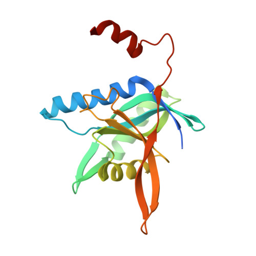
4QFT - PubMed Abstract:
The Cop9 signalosome complex (CSN) regulates the functional cycle of the major E3 ubiquitin ligase family, the cullin RING E3 ubiquitin ligases (CRLs). Activated CRLs are covalently modified by the ubiquitin-like protein Nedd8 (neural precursor cell expressed developmentally down-regulated protein 8). CSN serves an essential role in myriad cellular processes by reversing this modification through the isopeptidase activity of its CSN5 subunit. CSN5 alone is inactive due to an auto-inhibited conformation of its catalytic domain. Here we report the molecular basis of CSN5 catalytic domain activation and unravel a molecular hierarchy in CSN deneddylation activity. The association of CSN5 and CSN6 MPN (for Mpr1/Pad1 N-terminal) domains activates its isopeptidase activity. The CSN5/CSN6 module, however, is inefficient in CRL deneddylation, indicating a requirement of further elements in this reaction such as other CSN subunits. A hybrid molecular model of CSN5/CSN6 provides a structural framework to explain these functional observations. Docking this model into a published CSN electron density map and using distance constraints obtained from cross-linking coupled to mass-spectrometry, we find that the C-termini of the CSN subunits could form a helical bundle in the centre of the structure. They likely play a key scaffolding role in the spatial organization of CSN and precise positioning of the dimeric MPN catalytic core.
Organizational Affiliation:
Centre de Biochimie Structurale, Unité Mixte de Recherche (UMR) 5048, Centre National de Recherche Scientifique (CNRS), Université Montpellier 1 (UM1), Université Montpellier 2 (UM2), Montpellier, France; Institut national de la santé et de la recherche médicale (INSERM) U1054, Paris, France.