


Crystal structure of Vanderwaltozyma polyspora Nup133 Beta-propeller domain
Sampathkumar, P., Bonanno, J.B., Rout, M.P., Almo, S.C.To be published.
Experimental Data Snapshot
wwPDB Validation 3D Report Full Report

Entity ID: 1 | |||||
|---|---|---|---|---|---|
| Molecule | Chains | Sequence Length | Organism | Details | Image |
| Nucleoporin NUP133 | 459 | Vanderwaltozyma polyspora DSM 70294 | Mutation(s): 0 Gene Names: Kpol_1018p77, Nup133 | 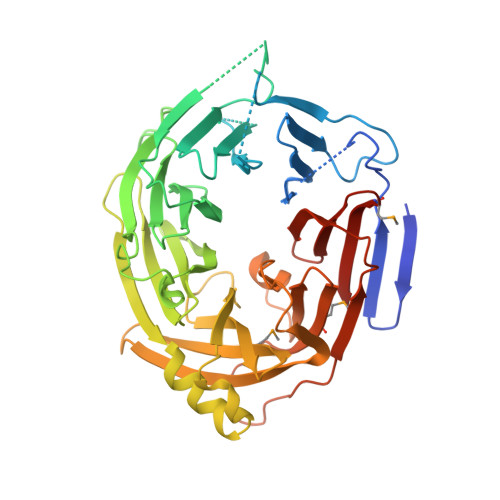 | |
UniProt | |||||
Find proteins for A7TDS5 (Vanderwaltozyma polyspora (strain ATCC 22028 / DSM 70294 / BCRC 21397 / CBS 2163 / NBRC 10782 / NRRL Y-8283 / UCD 57-17)) Explore A7TDS5 Go to UniProtKB: A7TDS5 | |||||
Entity Groups | |||||
| Sequence Clusters | 30% Identity50% Identity70% Identity90% Identity95% Identity100% Identity | ||||
| UniProt Group | A7TDS5 | ||||
Sequence AnnotationsExpand | |||||
| |||||
| Modified Residues 1 Unique | |||||
|---|---|---|---|---|---|
| ID | Chains | Type | Formula | 2D Diagram | Parent |
| MSE Query on MSE | A, B | L-PEPTIDE LINKING | C5 H11 N O2 Se |  | MET |
| Length ( Å ) | Angle ( ˚ ) |
|---|---|
| a = 109.172 | α = 90 |
| b = 133.843 | β = 90 |
| c = 136.775 | γ = 90 |
| Software Name | Purpose |
|---|---|
| REFMAC | refinement |
| PDB_EXTRACT | data extraction |
| XDS | data reduction |
| XDS | data scaling |
| Aimless | data scaling |
| PHENIX | phasing |
RCSB PDB (citation) is hosted by
RCSB PDB is a member of the

