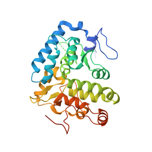
A conserved acidic residue in phenylalanine hydroxylase contributes to cofactor affinity and catalysis.
Ronau, J.A., Paul, L.N., Fuchs, J.E., Liedl, K.R., Abu-Omar, M.M., Das, C.(2014) Biochemistry 53: 6834-6848
- PubMed: 25295853
- DOI: https://doi.org/10.1021/bi500734h
- Primary Citation of Related Structures:
4Q3W, 4Q3X, 4Q3Y, 4Q3Z - PubMed Abstract:
The catalytic domains of aromatic amino acid hydroxylases (AAAHs) contain a non-heme iron coordinated to a 2-His-1-carboxylate facial triad and two water molecules. Asp139 from Chromobacterium violaceum PAH (cPAH) resides within the second coordination sphere and contributes key hydrogen bonds with three active site waters that mediate its interaction with an oxidized form of the cofactor, 7,8-dihydro-l-biopterin, in crystal structures. To determine the catalytic role of this residue, various point mutants were prepared and characterized. Our isothermal titration calorimetry (ITC) analysis of iron binding implies that polarity at position 139 is not the sole criterion for metal affinity, as binding studies with D139E suggest that the size of the amino acid side chain also appears to be important. High-resolution crystal structures of the mutants reveal that Asp139 may not be essential for holding the bridging water molecules together, because many of these waters are retained even in the Ala mutant. However, interactions via the bridging waters contribute to cofactor binding at the active site, interactions for which charge of the residue is important, as the D139N mutant shows a 5-fold decrease in its affinity for pterin as revealed by ITC (compared to a 16-fold loss of affinity in the case of the Ala mutant). The Asn and Ala mutants show a much more pronounced defect in their kcat values, with nearly 16- and 100-fold changes relative to that of the wild type, respectively, indicating a substantial role of this residue in stabilization of the transition state by aligning the cofactor in a productive orientation, most likely through direct binding with the cofactor, supported by data from molecular dynamics simulations of the complexes. Our results indicate that the intervening water structure between the cofactor and the acidic residue masks direct interaction between the two, possibly to prevent uncoupled hydroxylation of the cofactor before the arrival of phenylalanine. It thus appears that the second-coordination sphere Asp residue in cPAH, and, by extrapolation, the equivalent residue in other AAAHs, plays a role in fine-tuning pterin affinity in the ground state via deformable interactions with bridging waters and assumes a more significant role in the transition state by aligning the cofactor through direct hydrogen bonding.
Organizational Affiliation:
Department of Chemistry, Purdue University , 560 Oval Drive, West Lafayette, Indiana 47907, United States.