4PTF

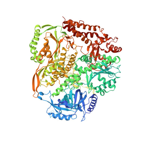
Ternary crystal structure of yeast DNA polymerase epsilon with template G


Experimental Data Snapshot

  • Method: X-RAY DIFFRACTION
  • Resolution: 2.81 Å
  • R-Value Free: 0.247 
  • R-Value Work: 0.183 
  • R-Value Observed: 0.186 

wwPDB Validation   3D Report Full Report


Ligand Structure Quality Assessment 


This is version 1.1 of the entry. See complete history


Literature

Crystal Structure of Yeast DNA Polymerase epsilon Catalytic Domain.

Jain, R.Rajashankar, K.R.Buku, A.Johnson, R.E.Prakash, L.Prakash, S.Aggarwal, A.K.

(2014) PLoS One 9: e94835-e94835

  • DOI: https://doi.org/10.1371/journal.pone.0094835
  • Primary Citation of Related Structures:  
    4PTF

  • PubMed Abstract: 

    DNA polymerase ε (Polε) is a multi-subunit polymerase that contributes to genomic stability via its roles in leading strand replication and the repair of damaged DNA. Here we report the ternary structure of the Polε catalytic subunit (Pol2) bound to a nascent G:C base pair (Pol2G:C). Pol2G:C has a typical B-family polymerase fold and embraces the template-primer duplex with the palm, fingers, thumb and exonuclease domains. The overall arrangement of domains is similar to the structure of Pol2T:A reported recently, but there are notable differences in their polymerase and exonuclease active sites. In particular, we observe Ca2+ ions at both positions A and B in the polymerase active site and also observe a Ca2+ at position B of the exonuclease site. We find that the contacts to the nascent G:C base pair in the Pol2G:C structure are maintained in the Pol2T:A structure and reflect the comparable fidelity of Pol2 for nascent purine-pyrimidine and pyrimidine-purine base pairs. We note that unlike that of Pol3, the shape of the nascent base pair binding pocket in Pol2 is modulated from the major grove side by the presence of Tyr431. Together with Pol2T:A, our results provide a framework for understanding the structural basis of high fidelity DNA synthesis by Pol2.


  • Organizational Affiliation

    Department of Structural and Chemical Biology, Mount Sinai School of Medicine, New York, New York, United States of America.


Macromolecules

Find similar proteins by:  (by identity cutoff)  |  3D Structure
Entity ID: 1
MoleculeChains Sequence LengthOrganismDetailsImage
DNA polymerase epsilon catalytic subunit A1,194Saccharomyces cerevisiaeMutation(s): 0 
Gene Names: POL2DUN2YNL262WN0825
EC: 2.7.7.7
UniProt
Find proteins for P21951 (Saccharomyces cerevisiae (strain ATCC 204508 / S288c))
Explore P21951 
Go to UniProtKB:  P21951
Entity Groups  
Sequence Clusters30% Identity50% Identity70% Identity90% Identity95% Identity100% Identity
UniProt GroupP21951
Sequence Annotations
Expand
  • Reference Sequence

Find similar nucleic acids by:  Sequence   |   3D Structure  

Entity ID: 2
MoleculeChains LengthOrganismImage
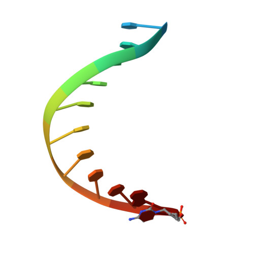
5'-D(*AP*TP*CP*CP*TP*CP*CP*CP*CP*TP*AP*(DOC))-3'B [auth P]12N/A
Sequence Annotations
Expand
  • Reference Sequence

Find similar nucleic acids by:  Sequence   |   3D Structure  

Entity ID: 3
MoleculeChains LengthOrganismImage
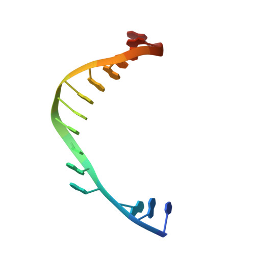
5'-D(*TP*AP*AP*GP*GP*TP*AP*GP*GP*GP*GP*AP*GP*GP*AP*T)-3'C [auth T]16N/A
Sequence Annotations
Expand
  • Reference Sequence
Small Molecules
Ligands 4 Unique
IDChains Name / Formula / InChI Key2D Diagram3D Interactions
DCP
Query on DCP

Download Ideal Coordinates CCD File 
D [auth A]2'-DEOXYCYTIDINE-5'-TRIPHOSPHATE
C9 H16 N3 O13 P3
RGWHQCVHVJXOKC-SHYZEUOFSA-N
EDO
Query on EDO

Download Ideal Coordinates CCD File 
J [auth T]1,2-ETHANEDIOL
C2 H6 O2
LYCAIKOWRPUZTN-UHFFFAOYSA-N
CA
Query on CA

Download Ideal Coordinates CCD File 
E [auth A],
F [auth A],
G [auth A],
I [auth T]
CALCIUM ION
Ca
BHPQYMZQTOCNFJ-UHFFFAOYSA-N
NA
Query on NA

Download Ideal Coordinates CCD File 
H [auth A]SODIUM ION
Na
FKNQFGJONOIPTF-UHFFFAOYSA-N
Experimental Data & Validation

Experimental Data

  • Method: X-RAY DIFFRACTION
  • Resolution: 2.81 Å
  • R-Value Free: 0.247 
  • R-Value Work: 0.183 
  • R-Value Observed: 0.186 
  • Space Group: C 1 2 1
Unit Cell:
Length ( Å )Angle ( ˚ )
a = 147.293α = 90
b = 68.478β = 109.6
c = 149.079γ = 90
Software Package:
Software NamePurpose
PHENIXrefinement
HKL-2000data reduction
HKL-2000data scaling
PHASERphasing

Structure Validation

View Full Validation Report


Ligand Structure Quality Assessment 


Entry History 

Deposition Data

Revision History  (Full details and data files)

  • Version 1.0: 2014-04-30
    Type: Initial release
  • Version 1.1: 2023-09-20
    Changes: Data collection, Database references, Derived calculations, Refinement description