


Crystal structure of POB3 middle domain at 1.65A
Liu, H.To be published.
Experimental Data Snapshot
wwPDB Validation 3D Report Full Report

Entity ID: 1 | |||||
|---|---|---|---|---|---|
| Molecule | Chains | Sequence Length | Organism | Details | Image |
| FACT complex subunit POB3 | 242 | Saccharomyces cerevisiae S288C | Mutation(s): 0 Gene Names: POB3, YML069W | 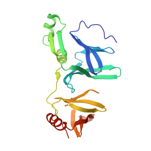 | |
UniProt | |||||
Find proteins for Q04636 (Saccharomyces cerevisiae (strain ATCC 204508 / S288c)) Explore Q04636 Go to UniProtKB: Q04636 | |||||
Entity Groups | |||||
| Sequence Clusters | 30% Identity50% Identity70% Identity90% Identity95% Identity100% Identity | ||||
| UniProt Group | Q04636 | ||||
Sequence AnnotationsExpand | |||||
| |||||
| Ligands 1 Unique | |||||
|---|---|---|---|---|---|
| ID | Chains | Name / Formula / InChI Key | 2D Diagram | 3D Interactions | |
| MLA Query on MLA | B [auth A] | MALONIC ACID C3 H4 O4 OFOBLEOULBTSOW-UHFFFAOYSA-N |  | ||
| Length ( Å ) | Angle ( ˚ ) |
|---|---|
| a = 31.08 | α = 90 |
| b = 59.15 | β = 90 |
| c = 144.93 | γ = 90 |
| Software Name | Purpose |
|---|---|
| HKL-2000 | data collection |
| AMoRE | phasing |
| REFMAC | refinement |
| MOSFLM | data reduction |
| SCALA | data scaling |
RCSB PDB (citation) is hosted by
RCSB PDB is a member of the

