


Crystal structure of 3-hydroxyacyl-CoA-dehydrogenase from Brucella melitensis
Lukacs, C.M., Abendroth, J., Edwards, T.E., Lorimer, D.To be published.
Experimental Data Snapshot
wwPDB Validation 3D Report Full Report

Entity ID: 1 | |||||
|---|---|---|---|---|---|
| Molecule | Chains | Sequence Length | Organism | Details | Image |
| 3-hydroxyacyl-CoA dehydrogenase | 263 | Brucella melitensis 64/150 | Mutation(s): 0 Gene Names: C045_02890 | 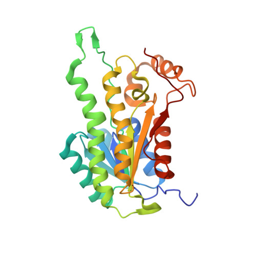 | |
Entity Groups | |||||
| Sequence Clusters | 30% Identity50% Identity70% Identity90% Identity95% Identity100% Identity | ||||
Sequence AnnotationsExpand | |||||
| |||||
| Ligands 1 Unique | |||||
|---|---|---|---|---|---|
| ID | Chains | Name / Formula / InChI Key | 2D Diagram | 3D Interactions | |
| TRS Query on TRS | I [auth B], J [auth D], K [auth F], L [auth G] | 2-AMINO-2-HYDROXYMETHYL-PROPANE-1,3-DIOL C4 H12 N O3 LENZDBCJOHFCAS-UHFFFAOYSA-O |  | ||
| Length ( Å ) | Angle ( ˚ ) |
|---|---|
| a = 72.84 | α = 90 |
| b = 72.84 | β = 90 |
| c = 350.41 | γ = 90 |
| Software Name | Purpose |
|---|---|
| REFMAC | refinement |
RCSB PDB (citation) is hosted by
RCSB PDB is a member of the

