4PFO

Myosin VI motor domain in the Pi release state, space group P212121


Experimental Data Snapshot

  • Method: X-RAY DIFFRACTION
  • Resolution: 1.75 Å
  • R-Value Free: 0.192 
  • R-Value Work: 0.169 
  • R-Value Observed: 0.171 

wwPDB Validation   3D Report Full Report


Ligand Structure Quality Assessment 


This is version 2.1 of the entry. See complete history


Literature

How actin initiates the motor activity of Myosin.

Llinas, P.Isabet, T.Song, L.Ropars, V.Zong, B.Benisty, H.Sirigu, S.Morris, C.Kikuti, C.Safer, D.Sweeney, H.L.Houdusse, A.

(2015) Dev Cell 33: 401-412

  • DOI: https://doi.org/10.1016/j.devcel.2015.03.025
  • Primary Citation of Related Structures:  
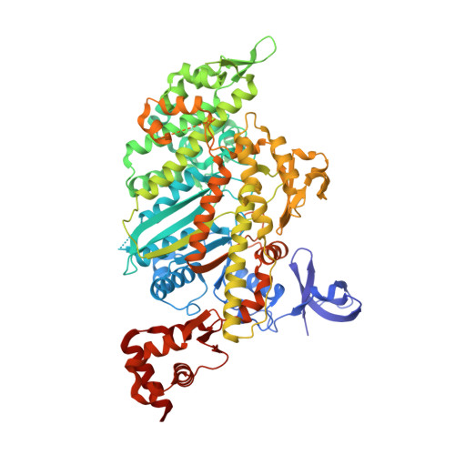
    4PFO, 4PFP, 4PJJ, 4PJK, 4PJL, 4PJM, 4PJN, 4PK4

  • PubMed Abstract: 

    Fundamental to cellular processes are directional movements driven by molecular motors. A common theme for these and other molecular machines driven by ATP is that controlled release of hydrolysis products is essential for using the chemical energy efficiently. Mechanochemical transduction by myosin motors on actin is coupled to unknown structural changes that result in the sequential release of inorganic phosphate (Pi) and MgADP. We present here a myosin structure possessing an actin-binding interface and a tunnel (back door) that creates an escape route for Pi with a minimal rotation of the myosin lever arm that drives movements. We propose that this state represents the beginning of the powerstroke on actin and that Pi translocation from the nucleotide pocket triggered by actin binding initiates myosin force generation. This elucidates how actin initiates force generation and movement and may represent a strategy common to many molecular machines.


  • Organizational Affiliation

    Structural Motility, Institut Curie, Centre de Recherche, Paris 75248, France; CNRS, UMR144, 26 rue d'Ulm, Cedex 05, Paris 75248, France.


Macromolecules
Find similar proteins by:  (by identity cutoff)  |  3D Structure
Entity ID: 1
MoleculeChains Sequence LengthOrganismDetailsImage
Unconventional myosin-VI788Sus scrofaMutation(s): 0 
Gene Names: MYO6
UniProt
Find proteins for F1RQI7 (Sus scrofa)
Explore F1RQI7 
Go to UniProtKB:  F1RQI7
Entity Groups  
Sequence Clusters30% Identity50% Identity70% Identity90% Identity95% Identity100% Identity
UniProt GroupF1RQI7
Sequence Annotations
Expand
  • Reference Sequence
Small Molecules
Ligands 3 Unique
IDChains Name / Formula / InChI Key2D Diagram3D Interactions
ADP
Query on ADP

Download Ideal Coordinates CCD File 
C [auth A]ADENOSINE-5'-DIPHOSPHATE
C10 H15 N5 O10 P2
XTWYTFMLZFPYCI-KQYNXXCUSA-N
GOL
Query on GOL

Download Ideal Coordinates CCD File 
D [auth A]
E [auth A]
F [auth A]
G [auth A]
H [auth A]
D [auth A],
E [auth A],
F [auth A],
G [auth A],
H [auth A],
I [auth A],
J [auth A],
K [auth A],
L [auth A],
M [auth A],
N [auth A],
O [auth A],
P [auth A],
Q [auth A]
GLYCEROL
C3 H8 O3
PEDCQBHIVMGVHV-UHFFFAOYSA-N
MG
Query on MG

Download Ideal Coordinates CCD File 
B [auth A]MAGNESIUM ION
Mg
JLVVSXFLKOJNIY-UHFFFAOYSA-N
Experimental Data & Validation

Experimental Data

  • Method: X-RAY DIFFRACTION
  • Resolution: 1.75 Å
  • R-Value Free: 0.192 
  • R-Value Work: 0.169 
  • R-Value Observed: 0.171 
  • Space Group: P 21 21 21
Unit Cell:
Length ( Å )Angle ( ˚ )
a = 91.27α = 90
b = 95.31β = 90
c = 103.52γ = 90
Software Package:
Software NamePurpose
BUSTERrefinement
Cootmodel building
PHASERphasing
XDSdata reduction
XSCALEdata scaling

Structure Validation

View Full Validation Report


Ligand Structure Quality Assessment 


Entry History & Funding Information

Deposition Data


Funding OrganizationLocationGrant Number
French National Research AgencyFranceBLAN07
French National Research AgencyFranceBLAN10
National Institutes of HealthUnited StatesRO01DC009100

Revision History  (Full details and data files)

  • Version 1.0: 2015-04-29
    Type: Initial release
  • Version 1.1: 2016-03-30
    Changes: Database references
  • Version 2.0: 2017-08-30
    Changes: Advisory, Atomic model, Author supporting evidence, Derived calculations
  • Version 2.1: 2023-12-20
    Changes: Data collection, Database references, Refinement description