4PC7

Elongation factor Tu:Ts complex in a near GTP conformation.


Experimental Data Snapshot

  • Method: X-RAY DIFFRACTION
  • Resolution: 3.60 Å
  • R-Value Free: 0.283 
  • R-Value Work: 0.255 
  • R-Value Observed: 0.257 

wwPDB Validation   3D Report Full Report


Ligand Structure Quality Assessment 


This is version 1.4 of the entry. See complete history


Literature

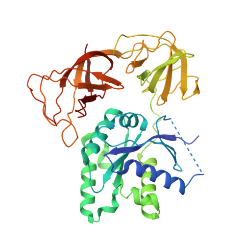
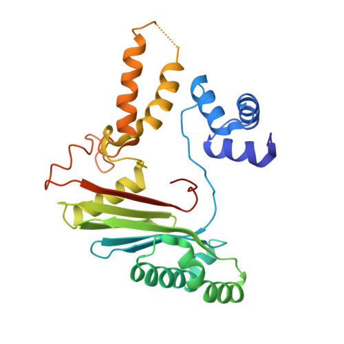
Structural outline of the detailed mechanism for elongation factor Ts-mediated guanine nucleotide exchange on elongation factor Tu.

Thirup, S.S.Van, L.B.Nielsen, T.K.Knudsen, C.R.

(2015) J Struct Biol 191: 10-21

  • DOI: https://doi.org/10.1016/j.jsb.2015.06.011
  • Primary Citation of Related Structures:  
    4PC1, 4PC2, 4PC3, 4PC6, 4PC7

  • PubMed Abstract: 

    Translation elongation factor EF-Tu belongs to the superfamily of guanine-nucleotide binding proteins, which play key cellular roles as regulatory switches. All G-proteins require activation via exchange of GDP for GTP to carry out their respective tasks. Often, guanine-nucleotide exchange factors are essential to this process. During translation, EF-Tu:GTP transports aminoacylated tRNA to the ribosome. GTP is hydrolyzed during this process, and subsequent reactivation of EF-Tu is catalyzed by EF-Ts. The reaction path of guanine-nucleotide exchange is structurally poorly defined for EF-Tu and EF-Ts. We have determined the crystal structures of the following reaction intermediates: two structures of EF-Tu:GDP:EF-Ts (2.2 and 1.8Å resolution), EF-Tu:PO4:EF-Ts (1.9Å resolution), EF-Tu:GDPNP:EF-Ts (2.2Å resolution) and EF-Tu:GDPNP:pulvomycin:Mg(2+):EF-Ts (3.5Å resolution). These structures provide snapshots throughout the entire exchange reaction and suggest a mechanism for the release of EF-Tu in its GTP conformation. An inferred sequence of events during the exchange reaction is presented.


  • Organizational Affiliation

    Aarhus University, Department of Molecular Biology and Genetics, Center for Structural Biology, DK-8000 Aarhus C, Denmark. Electronic address: sth@mbg.au.dk.


Macromolecules
Find similar proteins by:  (by identity cutoff)  |  3D Structure
Entity ID: 1
MoleculeChains Sequence LengthOrganismDetailsImage
Elongation factor Tu 1394Escherichia coli K-12Mutation(s): 0 
Gene Names: tufAb3339JW3301
UniProt
Find proteins for P0CE47 (Escherichia coli (strain K12))
Explore P0CE47 
Go to UniProtKB:  P0CE47
Entity Groups  
Sequence Clusters30% Identity50% Identity70% Identity90% Identity95% Identity100% Identity
UniProt GroupP0CE47
Sequence Annotations
Expand
  • Reference Sequence
Find similar proteins by:  (by identity cutoff)  |  3D Structure
Entity ID: 2
MoleculeChains Sequence LengthOrganismDetailsImage
Elongation factor TsB [auth C]282Escherichia coli K-12Mutation(s): 0 
Gene Names: tsfb0170JW0165
UniProt
Find proteins for P0A6P1 (Escherichia coli (strain K12))
Explore P0A6P1 
Go to UniProtKB:  P0A6P1
Entity Groups  
Sequence Clusters30% Identity50% Identity70% Identity90% Identity95% Identity100% Identity
UniProt GroupP0A6P1
Sequence Annotations
Expand
  • Reference Sequence
Small Molecules
Ligands 3 Unique
IDChains Name / Formula / InChI Key2D Diagram3D Interactions
PUL
Query on PUL

Download Ideal Coordinates CCD File 
D [auth A](1S,2S,3E,5E,7E,10S,11S,12S)-12-[(2R,4E,6E,8Z,10R,12E,14E,16Z,18S,19Z)-10,18-DIHYDROXY-12,16,19-TRIMETHYL-11,22-DIOXOOX ACYCLODOCOSA-4,6,8,12,14,16,19-HEPTAEN-2-YL]-2,11-DIHYDROXY-1,10-DIMETHYL-9-OXOTRIDECA-3,5,7-TRIEN-1-YL 6-DEOXY-2,4-DI-O-METHYL-BETA-L-GALACTOPYRANOSIDE
C47 H66 O13
FXSFWUNCIOIMAC-VXILMCRLSA-N
GNP
Query on GNP

Download Ideal Coordinates CCD File 
C [auth A]PHOSPHOAMINOPHOSPHONIC ACID-GUANYLATE ESTER
C10 H17 N6 O13 P3
UQABYHGXWYXDTK-UUOKFMHZSA-N
MG
Query on MG

Download Ideal Coordinates CCD File 
E [auth A]MAGNESIUM ION
Mg
JLVVSXFLKOJNIY-UHFFFAOYSA-N
Experimental Data & Validation

Experimental Data

  • Method: X-RAY DIFFRACTION
  • Resolution: 3.60 Å
  • R-Value Free: 0.283 
  • R-Value Work: 0.255 
  • R-Value Observed: 0.257 
  • Space Group: P 32 2 1
Unit Cell:
Length ( Å )Angle ( ˚ )
a = 122.82α = 90
b = 122.82β = 90
c = 173.33γ = 120
Software Package:
Software NamePurpose
PHENIXrefinement

Structure Validation

View Full Validation Report


Ligand Structure Quality Assessment 


Entry History 

Deposition Data

  • Released Date: 2015-05-06 
  • Deposition Author(s): Thirup, S.S.

Revision History  (Full details and data files)

  • Version 1.0: 2015-05-06
    Type: Initial release
  • Version 1.1: 2015-07-01
    Changes: Database references
  • Version 1.2: 2015-07-15
    Changes: Database references
  • Version 1.3: 2018-03-07
    Changes: Database references, Derived calculations, Source and taxonomy
  • Version 1.4: 2023-12-27
    Changes: Data collection, Database references, Derived calculations, Experimental preparation, Structure summary