4P5W

Structure of the N- and C-terminal domain fusion of the human mitochondrial aspartate/glutamate carrier Citrin in the calcium-bound state


Experimental Data Snapshot

  • Method: X-RAY DIFFRACTION
  • Resolution: 2.40 Å
  • R-Value Free: 0.227 
  • R-Value Work: 0.193 
  • R-Value Observed: 0.195 

wwPDB Validation   3D Report Full Report


This is version 2.0 of the entry. See complete history


Literature

Calcium-induced conformational changes of the regulatory domain of human mitochondrial aspartate/glutamate carriers.

Thangaratnarajah, C.Ruprecht, J.J.Kunji, E.R.

(2014) Nat Commun 5: 5491-5491

  • DOI: https://doi.org/10.1038/ncomms6491
  • Primary Citation of Related Structures:  
    4P5W, 4P5X, 4P60

  • PubMed Abstract: 

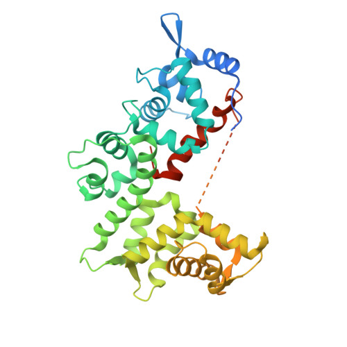
    The transport activity of human mitochondrial aspartate/glutamate carriers is central to the malate-aspartate shuttle, urea cycle, gluconeogenesis and myelin synthesis. They have a unique three-domain structure, comprising a calcium-regulated N-terminal domain with eight EF-hands, a mitochondrial carrier domain, and a C-terminal domain. Here we present the calcium-bound and calcium-free structures of the N- and C-terminal domains, elucidating the mechanism of calcium regulation. Unexpectedly, EF-hands 4-8 are involved in dimerization of the carrier and form a static unit, whereas EF-hands 1-3 form a calcium-responsive mobile unit. On calcium binding, an amphipathic helix of the C-terminal domain binds to the N-terminal domain, opening a vestibule. In the absence of calcium, the mobile unit closes the vestibule. Opening and closing of the vestibule might regulate access of substrates to the carrier domain, which is involved in their transport. These structures provide a framework for understanding cases of the mitochondrial disease citrin deficiency.


  • Organizational Affiliation

    The Medical Research Council, Mitochondrial Biology Unit, Wellcome Trust/MRC Building, Hills Road, Cambridge CB2 0XY, UK.


Macromolecules
Find similar proteins by:  (by identity cutoff)  |  3D Structure
Entity ID: 1
MoleculeChains Sequence LengthOrganismDetailsImage
Calcium-binding mitochondrial carrier protein Aralar2,Calcium-binding mitochondrial carrier protein Aralar2
A, B
411Homo sapiensMutation(s): 0 
Gene Names: SLC25A13ARALAR2
UniProt & NIH Common Fund Data Resources
Find proteins for Q9UJS0 (Homo sapiens)
Explore Q9UJS0 
Go to UniProtKB:  Q9UJS0
PHAROS:  Q9UJS0
GTEx:  ENSG00000004864 
Entity Groups  
Sequence Clusters30% Identity50% Identity70% Identity90% Identity95% Identity100% Identity
UniProt GroupQ9UJS0
Sequence Annotations
Expand
  • Reference Sequence
Experimental Data & Validation

Experimental Data

  • Method: X-RAY DIFFRACTION
  • Resolution: 2.40 Å
  • R-Value Free: 0.227 
  • R-Value Work: 0.193 
  • R-Value Observed: 0.195 
  • Space Group: P 21 21 21
Unit Cell:
Length ( Å )Angle ( ˚ )
a = 68.149α = 90
b = 106.167β = 90
c = 117.888γ = 90
Software Package:
Software NamePurpose
XDSdata reduction
Aimlessdata scaling
SHARPphasing
PHENIXrefinement

Structure Validation

View Full Validation Report


Entry History & Funding Information

Deposition Data


Funding OrganizationLocationGrant Number
Medical Research Council (United Kingdom)United Kingdom--
Mitochondrial European Educational TrainingGA n317433

Revision History  (Full details and data files)

  • Version 1.0: 2014-11-26
    Type: Initial release
  • Version 1.1: 2014-12-24
    Changes: Database references
  • Version 2.0: 2017-09-13
    Changes: Atomic model, Author supporting evidence