Proteolysis inside the Membrane Is a Rate-Governed Reaction Not Driven by Substrate Affinity.
Dickey, S.W., Baker, R.P., Cho, S., Urban, S.(2013) Cell 155: 1270-1281
- PubMed: 24315097
- DOI: https://doi.org/10.1016/j.cell.2013.10.053
- Primary Citation of Related Structures:
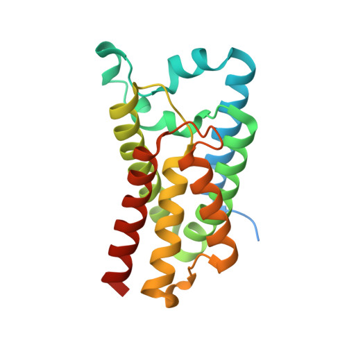
4NJN, 4NJP - PubMed Abstract:
Enzymatic cleavage of transmembrane anchors to release proteins from the membrane controls diverse signaling pathways and is implicated in more than a dozen diseases. How catalysis works within the viscous, water-excluding, two-dimensional membrane is unknown. We developed an inducible reconstitution system to interrogate rhomboid proteolysis quantitatively within the membrane in real time. Remarkably, rhomboid proteases displayed no physiological affinity for substrates (K(d) ~190 μM/0.1 mol%). Instead, ~10,000-fold differences in proteolytic efficiency with substrate mutants and diverse rhomboid proteases were reflected in k(cat) values alone. Analysis of gate-open mutant and solvent isotope effects revealed that substrate gating, not hydrolysis, is rate limiting. Ultimately, a single proteolytic event within the membrane normally takes minutes. Rhomboid intramembrane proteolysis is thus a slow, kinetically controlled reaction not driven by transmembrane protein-protein affinity. These properties are unlike those of other studied proteases or membrane proteins but are strikingly reminiscent of one subset of DNA-repair enzymes, raising important mechanistic and drug-design implications.
Organizational Affiliation:
Howard Hughes Medical Institute, Department of Molecular Biology & Genetics, Johns Hopkins University School of Medicine, Room 507 PCTB, 725 North Wolfe Street, Baltimore, MD 21205, USA.