4NBE

Fluorene-bound oxygenase with Phe275 replaced by Trp and ferredoxin complex of carbazole 1,9a-dioxygenase (form2)


Experimental Data Snapshot

  • Method: X-RAY DIFFRACTION
  • Resolution: 2.10 Å
  • R-Value Free: 0.252 
  • R-Value Work: 0.217 
  • R-Value Observed: 0.217 

wwPDB Validation   3D Report Full Report


Ligand Structure Quality Assessment 


This is version 1.2 of the entry. See complete history


Literature

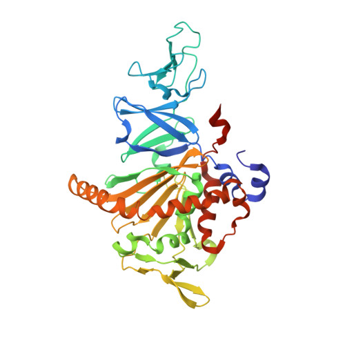
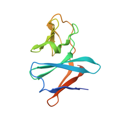
Structural basis of the divergent oxygenation reactions catalyzed by the rieske nonheme iron oxygenase carbazole 1,9a-dioxygenase.

Inoue, K.Usami, Y.Ashikawa, Y.Noguchi, H.Umeda, T.Yamagami-Ashikawa, A.Horisaki, T.Uchimura, H.Terada, T.Nakamura, S.Shimizu, K.Habe, H.Yamane, H.Fujimoto, Z.Nojiri, H.

(2014) Appl Environ Microbiol 80: 2821-2832

  • DOI: https://doi.org/10.1128/AEM.04000-13
  • Primary Citation of Related Structures:  
    4NB8, 4NB9, 4NBA, 4NBB, 4NBC, 4NBD, 4NBE, 4NBF, 4NBG, 4NBH

  • PubMed Abstract: 

    Carbazole 1,9a-dioxygenase (CARDO), a Rieske nonheme iron oxygenase (RO), is a three-component system composed of a terminal oxygenase (Oxy), ferredoxin, and a ferredoxin reductase. Oxy has angular dioxygenation activity against carbazole. Previously, site-directed mutagenesis of the Oxy-encoding gene from Janthinobacterium sp. strain J3 generated the I262V, F275W, Q282N, and Q282Y Oxy derivatives, which showed oxygenation capabilities different from those of the wild-type enzyme. To understand the structural features resulting in the different oxidation reactions, we determined the crystal structures of the derivatives, both free and complexed with substrates. The I262V, F275W, and Q282Y derivatives catalyze the lateral dioxygenation of carbazole with higher yields than the wild type. A previous study determined the crystal structure of Oxy complexed with carbazole and revealed that the carbonyl oxygen of Gly178 hydrogen bonds with the imino nitrogen of carbazole. In these derivatives, the carbazole was rotated approximately 15, 25, and 25°, respectively, compared to the wild type, creating space for a water molecule, which hydrogen bonds with the carbonyl oxygen of Gly178 and the imino nitrogen of carbazole. In the crystal structure of the F275W derivative complexed with fluorene, C-9 of fluorene, which corresponds to the imino nitrogen of carbazole, was oriented close to the mutated residue Trp275, which is on the opposite side of the binding pocket from the carbonyl oxygen of Gly178. Our structural analyses demonstrate that the fine-tuning of hydrophobic residues on the surface of the substrate-binding pocket in ROs causes a slight shift in the substrate-binding position that, in turn, favors specific oxygenation reactions toward various substrates.


  • Organizational Affiliation

    Biotechnology Research Center, The University of Tokyo, Bunkyo-ku, Tokyo, Japan.


Macromolecules
Find similar proteins by:  (by identity cutoff)  |  3D Structure
Entity ID: 1
MoleculeChains Sequence LengthOrganismDetailsImage
Terminal oxygenase component of carbazole
A, B, C
392Janthinobacterium sp. J3Mutation(s): 1 
Gene Names: carAa
EC: 1.14.12.22
UniProt
Find proteins for Q84II6 (Janthinobacterium sp. (strain J3))
Explore Q84II6 
Go to UniProtKB:  Q84II6
Entity Groups  
Sequence Clusters30% Identity50% Identity70% Identity90% Identity95% Identity100% Identity
UniProt GroupQ84II6
Sequence Annotations
Expand
  • Reference Sequence
Find similar proteins by:  (by identity cutoff)  |  3D Structure
Entity ID: 2
MoleculeChains Sequence LengthOrganismDetailsImage
Ferredoxin CarAc
D, E
115Pseudomonas resinovoransMutation(s): 0 
Gene Names: carAc
EC: 1.14.12.22
UniProt
Find proteins for Q8GI16 (Pseudomonas resinovorans)
Explore Q8GI16 
Go to UniProtKB:  Q8GI16
Entity Groups  
Sequence Clusters30% Identity50% Identity70% Identity90% Identity95% Identity100% Identity
UniProt GroupQ8GI16
Sequence Annotations
Expand
  • Reference Sequence
Small Molecules
Ligands 3 Unique
IDChains Name / Formula / InChI Key2D Diagram3D Interactions
FES
Query on FES

Download Ideal Coordinates CCD File 
G [auth A],
J [auth B],
L [auth C],
N [auth D],
O [auth E]
FE2/S2 (INORGANIC) CLUSTER
Fe2 S2
NIXDOXVAJZFRNF-UHFFFAOYSA-N
9FL
Query on 9FL

Download Ideal Coordinates CCD File 
H [auth A],
M [auth C]
9H-fluorene
C13 H10
NIHNNTQXNPWCJQ-UHFFFAOYSA-N
FE2
Query on FE2

Download Ideal Coordinates CCD File 
F [auth A],
I [auth B],
K [auth C]
FE (II) ION
Fe
CWYNVVGOOAEACU-UHFFFAOYSA-N
Experimental Data & Validation

Experimental Data

  • Method: X-RAY DIFFRACTION
  • Resolution: 2.10 Å
  • R-Value Free: 0.252 
  • R-Value Work: 0.217 
  • R-Value Observed: 0.217 
  • Space Group: P 1 21 1
Unit Cell:
Length ( Å )Angle ( ˚ )
a = 98.455α = 90
b = 88.264β = 106.72
c = 108.621γ = 90
Software Package:
Software NamePurpose
ADSCdata collection
MOLREPphasing
CNSrefinement
HKL-2000data reduction
HKL-2000data scaling

Structure Validation

View Full Validation Report


Ligand Structure Quality Assessment 


Entry History 

Deposition Data

Revision History  (Full details and data files)

  • Version 1.0: 2014-03-26
    Type: Initial release
  • Version 1.1: 2022-08-24
    Changes: Database references, Derived calculations
  • Version 1.2: 2023-11-08
    Changes: Data collection, Refinement description