4N8N

Crystal structure of Mycobacterial FtsX extracellular domain


Experimental Data Snapshot

  • Method: X-RAY DIFFRACTION
  • Resolution: 1.87 Å
  • R-Value Free: 0.233 
  • R-Value Work: 0.186 
  • R-Value Observed: 0.189 

wwPDB Validation   3D Report Full Report


This is version 1.3 of the entry. See complete history


Literature

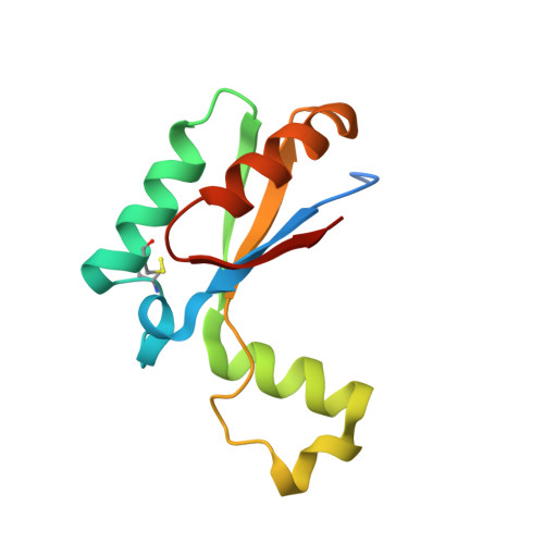
Mycobacterium tuberculosis FtsX extracellular domain activates the peptidoglycan hydrolase, RipC.

Mavrici, D.Marakalala, M.J.Holton, J.M.Prigozhin, D.M.Gee, C.L.Zhang, Y.J.Rubin, E.J.Alber, T.

(2014) Proc Natl Acad Sci U S A 111: 8037-8042

  • DOI: https://doi.org/10.1073/pnas.1321812111
  • Primary Citation of Related Structures:  
    4N8N, 4N8O

  • PubMed Abstract: 

    Bacterial growth and cell division are coordinated with hydrolysis of the peptidoglycan (PG) layer of the cell wall, but the mechanisms of regulation of extracellular PG hydrolases are not well understood. Here we report the biochemical, structural, and genetic analysis of the Mycobacterium tuberculosis homolog of the transmembrane PG-hydrolase regulator, FtsX. The purified FtsX extracellular domain binds the PG peptidase Rv2190c/RipC N-terminal segment, causing a conformational change that activates the enzyme. Deletion of ftsEX and ripC caused similar phenotypes in Mycobacterium smegmatis, as expected for genes in a single pathway. The crystal structure of the FtsX extracellular domain reveals an unprecedented fold containing two lobes connected by a flexible hinge. Mutations in the hydrophobic cleft between the lobes reduce RipC binding in vitro and inhibit FtsX function in M. smegmatis. These studies suggest how FtsX recognizes RipC and support a model in which a conformational change in FtsX links the cell division apparatus with PG hydrolysis.


  • Organizational Affiliation

    QB3 Institute, California Institute for Quantitative Bioscience, and.


Macromolecules
Find similar proteins by:  (by identity cutoff)  |  3D Structure
Entity ID: 1
MoleculeChains Sequence LengthOrganismDetailsImage
Cell division protein FtsX
A, B, C, D
115Mycobacterium tuberculosis H37RvMutation(s): 0 
Gene Names: ftsXMT3185MTCY164.12cRv3101c
UniProt
Find proteins for P9WG19 (Mycobacterium tuberculosis (strain ATCC 25618 / H37Rv))
Explore P9WG19 
Go to UniProtKB:  P9WG19
Entity Groups  
Sequence Clusters30% Identity50% Identity70% Identity90% Identity95% Identity100% Identity
UniProt GroupP9WG19
Sequence Annotations
Expand
  • Reference Sequence
Experimental Data & Validation

Experimental Data

  • Method: X-RAY DIFFRACTION
  • Resolution: 1.87 Å
  • R-Value Free: 0.233 
  • R-Value Work: 0.186 
  • R-Value Observed: 0.189 
  • Space Group: P 21 21 2
Unit Cell:
Length ( Å )Angle ( ˚ )
a = 106.248α = 90
b = 150.009β = 90
c = 28.658γ = 90
Software Package:
Software NamePurpose
DENZOdata reduction
SCALEPACKdata scaling
PHENIXrefinement
PDB_EXTRACTdata extraction
ADSCdata collection
HKL-2000data reduction
SOLVEphasing

Structure Validation

View Full Validation Report


Entry History 

Deposition Data

  • Released Date: 2014-05-28 
  • Deposition Author(s): Mavrici, D.

Revision History  (Full details and data files)

  • Version 1.0: 2014-05-28
    Type: Initial release
  • Version 1.1: 2014-07-09
    Changes: Database references
  • Version 1.2: 2014-07-16
    Changes: Structure summary
  • Version 1.3: 2017-11-15
    Changes: Refinement description