4MWS

Crystal structure of human PPCA (trigonal crystal form 1)


Experimental Data Snapshot

  • Method: X-RAY DIFFRACTION
  • Resolution: 2.80 Å
  • R-Value Free: 0.195 
  • R-Value Work: 0.146 
  • R-Value Observed: 0.149 

wwPDB Validation   3D Report Full Report


Ligand Structure Quality Assessment 


This is version 2.1 of the entry. See complete history


Literature

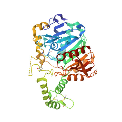
Proteolytic activation of human cathepsin A.

Kolli, N.Garman, S.C.

(2014) J Biol Chem 289: 11592-11600

  • DOI: https://doi.org/10.1074/jbc.M113.524280
  • Primary Citation of Related Structures:  
    4MWS, 4MWT

  • PubMed Abstract: 

    Galactosialidosis is a human lysosomal storage disease caused by deficiency in the multifunctional lysosomal protease cathepsin A (also known as protective protein/cathepsin A, PPCA, catA, HPP, and CTSA; EC 3.4.16.5). Previous structural work on the inactive precursor human cathepsin A (zymogen) led to a two-stage model for activation, where proteolysis of a 1.6-kDa excision peptide is followed by a conformational change in a blocking peptide occluding the active site. Here we present evidence for an alternate model of activation of human cathepsin A, needing only cleavage of a 3.3-kDa excision peptide to yield full enzymatic activity, with no conformational change required. We present x-ray crystallographic, mass spectrometric, amino acid sequencing, enzymatic, and cellular data to support the cleavage-only activation model. The results clarify a longstanding question about the mechanism of cathepsin A activation and point to new avenues for the design of mechanism-based inhibitors of the enzyme.


  • Organizational Affiliation

    Department of Biochemistry and Molecular Biology, University of Massachusetts, Amherst, Massachusetts 01003; Program in Molecular and Cellular Biology, University of Massachusetts, Amherst, Massachusetts 01003.


Macromolecules
Find similar proteins by:  (by identity cutoff)  |  3D Structure
Entity ID: 1
MoleculeChains Sequence LengthOrganismDetailsImage
Lysosomal protective protein
A, B
428Homo sapiensMutation(s): 0 
Gene Names: CTSAPPGB
EC: 3.4.16.5
UniProt & NIH Common Fund Data Resources
Find proteins for P10619 (Homo sapiens)
Explore P10619 
Go to UniProtKB:  P10619
PHAROS:  P10619
GTEx:  ENSG00000064601 
Entity Groups  
Sequence Clusters30% Identity50% Identity70% Identity90% Identity95% Identity100% Identity
UniProt GroupP10619
Sequence Annotations
Expand
  • Reference Sequence
Oligosaccharides

Help

Entity ID: 2
MoleculeChains Length2D Diagram Glycosylation3D Interactions
alpha-D-mannopyranose-(1-3)-beta-D-mannopyranose-(1-4)-2-acetamido-2-deoxy-beta-D-glucopyranose-(1-4)-[alpha-L-fucopyranose-(1-3)]2-acetamido-2-deoxy-beta-D-glucopyranose
C
5N-Glycosylation
Glycosylation Resources
GlyTouCan:  G17689EW
GlyCosmos:  G17689EW
GlyGen:  G17689EW
Entity ID: 3
MoleculeChains Length2D Diagram Glycosylation3D Interactions
beta-D-mannopyranose-(1-4)-2-acetamido-2-deoxy-beta-D-glucopyranose-(1-4)-[alpha-L-fucopyranose-(1-3)]2-acetamido-2-deoxy-beta-D-glucopyranose
D
4N-Glycosylation
Glycosylation Resources
GlyTouCan:  G18638YB
GlyCosmos:  G18638YB
GlyGen:  G18638YB
Experimental Data & Validation

Experimental Data

  • Method: X-RAY DIFFRACTION
  • Resolution: 2.80 Å
  • R-Value Free: 0.195 
  • R-Value Work: 0.146 
  • R-Value Observed: 0.149 
  • Space Group: P 31 2 1
Unit Cell:
Length ( Å )Angle ( ˚ )
a = 134.889α = 90
b = 134.889β = 90
c = 99.805γ = 120
Software Package:
Software NamePurpose
DENZOdata reduction
SCALEPACKdata scaling
PHASERphasing
REFMACrefinement
PDB_EXTRACTdata extraction

Structure Validation

View Full Validation Report


Ligand Structure Quality Assessment 


Entry History 

Deposition Data

Revision History  (Full details and data files)

  • Version 1.0: 2014-03-12
    Type: Initial release
  • Version 1.1: 2015-04-15
    Changes: Database references
  • Version 1.2: 2017-08-23
    Changes: Data collection
  • Version 2.0: 2020-07-29
    Type: Remediation
    Reason: Carbohydrate remediation
    Changes: Atomic model, Data collection, Database references, Derived calculations, Structure summary
  • Version 2.1: 2023-09-20
    Changes: Data collection, Database references, Refinement description, Structure summary