4MCQ

A high resolution structure of human glutamate carboxypeptidase II (GCPII) in complex with folyldi-gamma-L-glutamic acid (pteroyltri-gamma-L-glutamic acid)


Experimental Data Snapshot

  • Method: X-RAY DIFFRACTION
  • Resolution: 2.00 Å
  • R-Value Free: 0.172 
  • R-Value Work: 0.137 
  • R-Value Observed: 0.138 

wwPDB Validation   3D Report Full Report


Ligand Structure Quality Assessment 


This is version 2.0 of the entry. See complete history


Literature

Structural and biochemical characterization of the folyl-poly-gamma-l-glutamate hydrolyzing activity of human glutamate carboxypeptidase II.

Navratil, M.Ptacek, J.Sacha, P.Starkova, J.Lubkowski, J.Barinka, C.Konvalinka, J.

(2014) FEBS J 281: 3228-3242

  • DOI: https://doi.org/10.1111/febs.12857
  • Primary Citation of Related Structures:  
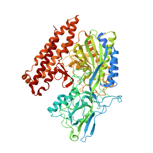
    4MCP, 4MCQ, 4MCR, 4MCS

  • PubMed Abstract: 

    In addition to its well-characterized role in the central nervous system, human glutamate carboxypeptidase II (GCPII; Uniprot ID Q04609) acts as a folate hydrolase in the small intestine, participating in the absorption of dietary polyglutamylated folates (folyl-n-γ-l-glutamic acid), which are the provitamin form of folic acid (also known as vitamin B9 ). Despite the role of GCPII as a folate hydrolase, nothing is known about the processing of polyglutamylated folates by GCPII at the structural or enzymological level. Moreover, many epidemiologic studies on the relationship of the naturally occurring His475Tyr polymorphism to folic acid status suggest that this polymorphism may be associated with several pathologies linked to impaired folate metabolism. In the present study, we report: (a) a series X-ray structures of complexes between a catalytically inactive GCPII mutant (Glu424Ala) and a panel of naturally occurring polyglutamylated folates; (b) the X-ray structure of the His475Tyr variant at a resolution of 1.83 Å; (c) the study of the recently identified arene-binding site of GCPII through mutagenesis (Arg463Leu, Arg511Leu and Trp541Ala), inhibitor binding and enzyme kinetics with polyglutamylated folates as substrates; and (d) a comparison of the thermal stabilities and folate-hydrolyzing activities of GCPII wild-type and His475Tyr variants. As a result, the crystallographic data reveal considerable details about the binding mode of polyglutamylated folates to GCPII, especially the engagement of the arene binding site in recognizing the folic acid moiety. Additionally, the combined structural and kinetic data suggest that GCPII wild-type and His475Tyr variant are functionally identical.


  • Organizational Affiliation

    Gilead Sciences and IOCB Research Centre, Institute of Organic Chemistry and Biochemistry, Academy of Sciences of the Czech Republic, Prague 6, Czech Republic; Department of Biochemistry, Faculty of Sciences, Charles University in Prague, Czech Republic.


Macromolecules
Find similar proteins by:  (by identity cutoff)  |  3D Structure
Entity ID: 1
MoleculeChains Sequence LengthOrganismDetailsImage
Glutamate carboxypeptidase 2757Homo sapiensMutation(s): 1 
Gene Names: FOLH1FOLHNAALAD1PSMPSMAGIG27
EC: 3.4.17.21
UniProt & NIH Common Fund Data Resources
Find proteins for Q04609 (Homo sapiens)
Explore Q04609 
Go to UniProtKB:  Q04609
PHAROS:  Q04609
GTEx:  ENSG00000086205 
Entity Groups  
Sequence Clusters30% Identity50% Identity70% Identity90% Identity95% Identity100% Identity
UniProt GroupQ04609
Sequence Annotations
Expand
  • Reference Sequence
Oligosaccharides

Help

Entity ID: 2
MoleculeChains Length2D Diagram Glycosylation3D Interactions
2-acetamido-2-deoxy-beta-D-glucopyranose-(1-4)-2-acetamido-2-deoxy-beta-D-glucopyranose
B, C, D
2N-Glycosylation
Glycosylation Resources
GlyTouCan:  G42666HT
GlyCosmos:  G42666HT
GlyGen:  G42666HT
Entity ID: 3
MoleculeChains Length2D Diagram Glycosylation3D Interactions
alpha-D-mannopyranose-(1-3)-beta-D-mannopyranose-(1-4)-2-acetamido-2-deoxy-beta-D-glucopyranose-(1-4)-2-acetamido-2-deoxy-beta-D-glucopyranose
E
4N-Glycosylation
Glycosylation Resources
GlyTouCan:  G81315DD
GlyCosmos:  G81315DD
GlyGen:  G81315DD
Small Molecules
Ligands 5 Unique
IDChains Name / Formula / InChI Key2D Diagram3D Interactions
29C
Query on 29C

Download Ideal Coordinates CCD File 
M [auth A]N-(4-{[(2-amino-4-oxo-3,4-dihydropteridin-6-yl)methyl]amino}benzoyl)-L-gamma-glutamyl-L-gamma-glutamyl-L-glutamic acid
C29 H33 N9 O12
WOLQREOUPKZMEX-BZSNNMDCSA-N
NAG
Query on NAG

Download Ideal Coordinates CCD File 
J [auth A],
K [auth A],
L [auth A]
2-acetamido-2-deoxy-beta-D-glucopyranose
C8 H15 N O6
OVRNDRQMDRJTHS-FMDGEEDCSA-N
ZN
Query on ZN

Download Ideal Coordinates CCD File 
F [auth A],
G [auth A]
ZINC ION
Zn
PTFCDOFLOPIGGS-UHFFFAOYSA-N
CA
Query on CA

Download Ideal Coordinates CCD File 
H [auth A]CALCIUM ION
Ca
BHPQYMZQTOCNFJ-UHFFFAOYSA-N
CL
Query on CL

Download Ideal Coordinates CCD File 
I [auth A]CHLORIDE ION
Cl
VEXZGXHMUGYJMC-UHFFFAOYSA-M
Experimental Data & Validation

Experimental Data

  • Method: X-RAY DIFFRACTION
  • Resolution: 2.00 Å
  • R-Value Free: 0.172 
  • R-Value Work: 0.137 
  • R-Value Observed: 0.138 
  • Space Group: I 2 2 2
Unit Cell:
Length ( Å )Angle ( ˚ )
a = 101.646α = 90
b = 130.14β = 90
c = 159.867γ = 90
Software Package:
Software NamePurpose
BL-Controldata collection
REFMACrefinement
HKL-2000data reduction
HKL-2000data scaling
REFMACphasing

Structure Validation

View Full Validation Report


Ligand Structure Quality Assessment 


Entry History 

Deposition Data

Revision History  (Full details and data files)

  • Version 1.0: 2014-06-18
    Type: Initial release
  • Version 1.1: 2014-08-13
    Changes: Database references
  • Version 2.0: 2020-07-29
    Type: Remediation
    Reason: Carbohydrate remediation
    Changes: Advisory, Atomic model, Data collection, Database references, Derived calculations, Structure summary