


The high resolution structure of tyrocidine A reveals an amphipathic dimer.
Loll, P.J., Upton, E.C., Nahoum, V., Economou, N.J., Cocklin, S.(2014) Biochim Biophys Acta 1838: 1199-1207
- PubMed: 24530898
- DOI: https://doi.org/10.1016/j.bbamem.2014.01.033
- Primary Citation of Related Structures:
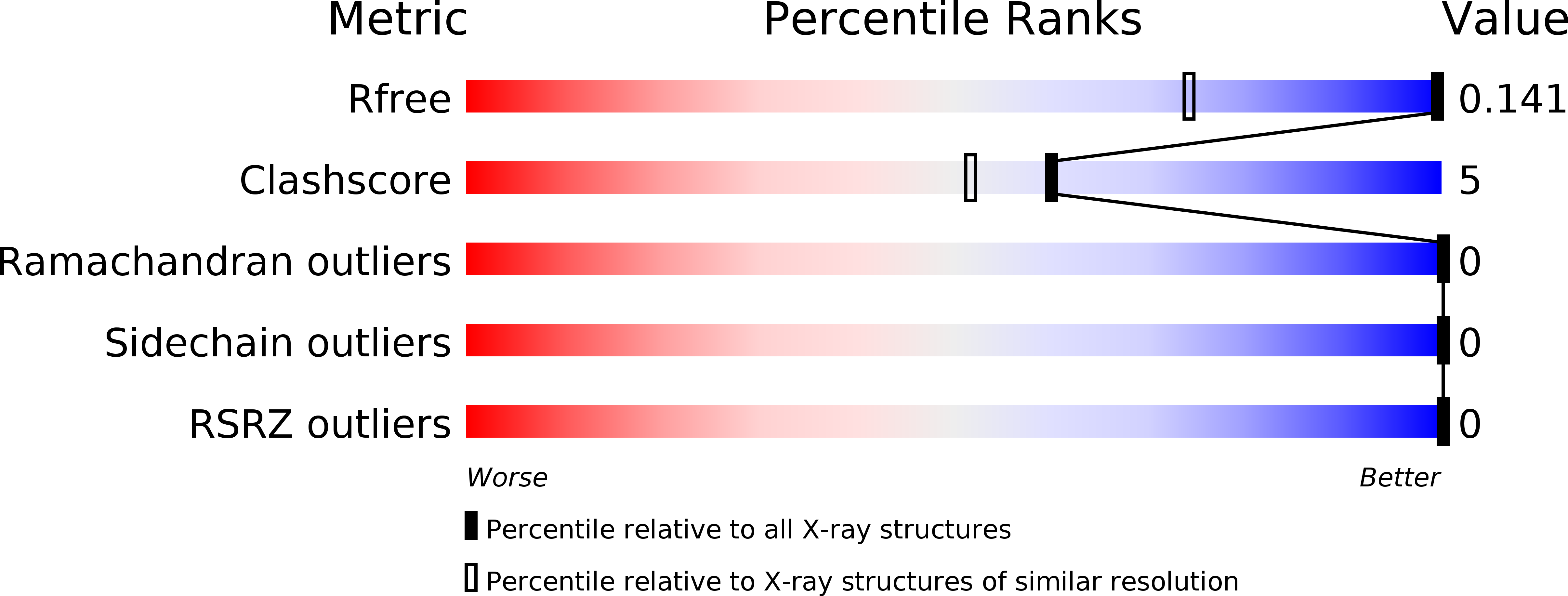
4M6E - PubMed Abstract:
Tyrocidine A, one of the first antibiotics ever to be discovered, is a cyclic decapeptide that binds to membranes of target bacteria, disrupting their integrity. It is active against a broad spectrum of Gram-positive organisms, and has recently engendered interest as a potential scaffold for the development of new drugs to combat antibiotic-resistant pathogens. We present here the X-ray crystal structure of tyrocidine A at a resolution of 0.95Å. The structure reveals that tyrocidine forms an intimate and highly amphipathic homodimer made up of four beta strands that associate into a single, highly curved antiparallel beta sheet. We used surface plasmon resonance and potassium efflux assays to demonstrate that tyrocidine binds tightly to mimetics of bacterial membranes with an apparent dissociation constant (K(D)) of 10 μM, and efficiently permeabilizes bacterial cells at concentrations equal to and below the K(D). Using variant forms of tyrocidine in which the fluorescent probe p-cyano-phenylalanine had been inserted on either the polar or apolar face of the molecule, we performed fluorescence quenching experiments, using both water-soluble and membrane-embedded quenchers. The quenching results, together with the structure, strongly support a membrane association model in which the convex, apolar face of tyrocidine's beta sheet is oriented toward the membrane interior, while the concave, polar face is presented to the aqueous phase.
Organizational Affiliation:
Department of Biochemistry and Molecular Biology, Drexel University College of Medicine, Philadelphia, PA 19102, USA. Electronic address: ploll@drexelmed.edu.


