4M1J

Crystal structure of Pseudomonas aeruginosa PvdQ in complex with a transition state analogue


Experimental Data Snapshot

  • Method: X-RAY DIFFRACTION
  • Resolution: 1.80 Å
  • R-Value Free: 0.188 
  • R-Value Work: 0.162 
  • R-Value Observed: 0.164 

wwPDB Validation   3D Report Full Report


Ligand Structure Quality Assessment 


This is version 1.2 of the entry. See complete history


Literature

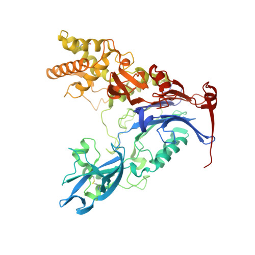
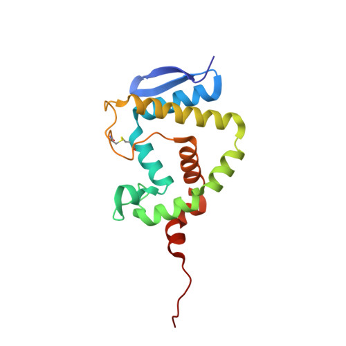
Rational Design of a Transition State Analogue with Picomolar Affinity for Pseudomonas aeruginosa PvdQ, a Siderophore Biosynthetic Enzyme.

Clevenger, K.D.Wu, R.Er, J.A.Liu, D.Fast, W.

(2013) ACS Chem Biol 8: 2192-2200

  • DOI: https://doi.org/10.1021/cb400345h
  • Primary Citation of Related Structures:  
    4M1J

  • PubMed Abstract: 

    The Pseudomonas aeruginosa enzyme PvdQ can process different substrates involved in quorum-sensing or in siderophore biosynthesis. Substrate selectivity was evaluated using steady-state kinetic constants for hydrolysis of N-acyl-homoserine lactones (HSLs) and p-nitrophenyl fatty acid esters. PvdQ prefers substrates with alkyl chains between 12 and 14 carbons long that do not bear a 3-oxo substitution and is revealed here to have a relatively high specificity constant for selected N-acyl-HSLs (kcat/KM = 10(5) to 10(6) M(-1) s(-1)). However, endogenous P. aeruginosa N-acyl-HSLs are ≥100-fold disfavored, supporting the conclusion that PvdQ was not primarily evolved to regulate endogenous quorum-sensing. PvdQ plays an essential biosynthetic role for the siderophore pyoverdine, on which P. aeruginosa depends for growth in iron-limited environments. A series of alkylboronate inhibitors was found to be reversible, competitive, and extremely potent (Ki ≥ 190 pM). A 1.8 Å X-ray structure shows that 1-tridecylboronic acid forms a monocovalent bond with the N-terminal β-chain Ser residue in the PvdQ heterodimer, mimicking a reaction transition state. This boronic acid inhibits growth of P. aeruginosa in iron-limited media, reproducing the phenotype of a genetic pvdQ disruption, although co-administration of an efflux pump inhibitor is required to maintain growth inhibition. These findings support the strategy of designing boron-based inhibitors of siderophore biosynthetic enzymes to control P. aeruginosa infections.


  • Organizational Affiliation

    Department of Chemistry and Biochemistry and the ‡College of Pharmacy, Medicinal Chemistry Division, University of Texas , Austin, Texas 78712, United States.


Macromolecules
Find similar proteins by:  (by identity cutoff)  |  3D Structure
Entity ID: 1
MoleculeChains Sequence LengthOrganismDetailsImage
Acyl-homoserine lactone acylase PvdQ subunit betaA [auth C]548Pseudomonas aeruginosaMutation(s): 0 
Gene Names: PA2385pvdQqsc112
EC: 3.5.1.97
UniProt
Find proteins for Q9I194 (Pseudomonas aeruginosa (strain ATCC 15692 / DSM 22644 / CIP 104116 / JCM 14847 / LMG 12228 / 1C / PRS 101 / PAO1))
Explore Q9I194 
Go to UniProtKB:  Q9I194
Entity Groups  
Sequence Clusters30% Identity50% Identity70% Identity90% Identity95% Identity100% Identity
UniProt GroupQ9I194
Sequence Annotations
Expand
  • Reference Sequence
Find similar proteins by:  (by identity cutoff)  |  3D Structure
Entity ID: 2
MoleculeChains Sequence LengthOrganismDetailsImage
Acyl-homoserine lactone acylase PvdQ subunit alphaB [auth A]165Pseudomonas aeruginosaMutation(s): 0 
Gene Names: pvdQqsc112PA2385
EC: 3.5.1.97
UniProt
Find proteins for Q9I194 (Pseudomonas aeruginosa (strain ATCC 15692 / DSM 22644 / CIP 104116 / JCM 14847 / LMG 12228 / 1C / PRS 101 / PAO1))
Explore Q9I194 
Go to UniProtKB:  Q9I194
Entity Groups  
Sequence Clusters30% Identity50% Identity70% Identity90% Identity95% Identity100% Identity
UniProt GroupQ9I194
Sequence Annotations
Expand
  • Reference Sequence
Small Molecules
Ligands 2 Unique
IDChains Name / Formula / InChI Key2D Diagram3D Interactions
B0S
Query on B0S

Download Ideal Coordinates CCD File 
C
tridecylboronic acid
C13 H29 B O2
IBKXABQGVIIFFS-UHFFFAOYSA-N
GOL
Query on GOL

Download Ideal Coordinates CCD File 
D [auth C]
E [auth C]
F [auth C]
G [auth C]
H [auth C]
D [auth C],
E [auth C],
F [auth C],
G [auth C],
H [auth C],
I [auth C],
J [auth C],
K [auth C],
L [auth C],
M [auth C],
N [auth C],
O [auth C],
P [auth C],
Q [auth C],
R [auth A],
S [auth A],
T [auth A],
U [auth A],
V [auth A],
W [auth A]
GLYCEROL
C3 H8 O3
PEDCQBHIVMGVHV-UHFFFAOYSA-N
Binding Affinity Annotations 
IDSourceBinding Affinity
B0S PDBBind:  4M1J Ki: 0.2 (nM) from 1 assay(s)
Experimental Data & Validation

Experimental Data

  • Method: X-RAY DIFFRACTION
  • Resolution: 1.80 Å
  • R-Value Free: 0.188 
  • R-Value Work: 0.162 
  • R-Value Observed: 0.164 
  • Space Group: C 2 2 21
Unit Cell:
Length ( Å )Angle ( ˚ )
a = 120.948α = 90
b = 166.433β = 90
c = 94.208γ = 90
Software Package:
Software NamePurpose
SCALEPACKdata scaling
PHASERphasing
PHENIXrefinement
PDB_EXTRACTdata extraction
HKL-2000data collection
DENZOdata reduction

Structure Validation

View Full Validation Report


Ligand Structure Quality Assessment 


Entry History 

Deposition Data

Revision History  (Full details and data files)

  • Version 1.0: 2013-08-28
    Type: Initial release
  • Version 1.1: 2013-11-06
    Changes: Database references
  • Version 1.2: 2017-11-15
    Changes: Refinement description