4LVI

MobM Relaxase Domain (MOBV; Mob_Pre) bound to plasmid pMV158 oriT DNA (22nt). Mn-bound crystal structure at pH 4.6


Experimental Data Snapshot

  • Method: X-RAY DIFFRACTION
  • Resolution: 1.90 Å
  • R-Value Free: 0.248 
  • R-Value Work: 0.206 
  • R-Value Observed: 0.208 

wwPDB Validation   3D Report Full Report


This is version 1.2 of the entry. See complete history


Literature

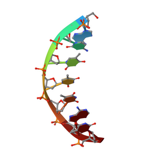
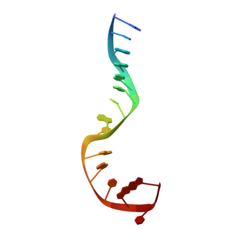
Structural basis of a histidine-DNA nicking/joining mechanism for gene transfer and promiscuous spread of antibiotic resistance.

Pluta, R.Boer, D.R.Lorenzo-Diaz, F.Russi, S.Gomez, H.Fernandez-Lopez, C.Perez-Luque, R.Orozco, M.Espinosa, M.Coll, M.

(2017) Proc Natl Acad Sci U S A 114: E6526-E6535

  • DOI: https://doi.org/10.1073/pnas.1702971114
  • Primary Citation of Related Structures:  
    4LVI, 4LVJ, 4LVK, 4LVL, 4LVM, 5N2Q

  • PubMed Abstract: 

    Relaxases are metal-dependent nucleases that break and join DNA for the initiation and completion of conjugative bacterial gene transfer. Conjugation is the main process through which antibiotic resistance spreads among bacteria, with multidrug-resistant staphylococci and streptococci infections posing major threats to human health. The MOB V family of relaxases accounts for approximately 85% of all relaxases found in Staphylococcus aureus isolates. Here, we present six structures of the MOB V relaxase MobM from the promiscuous plasmid pMV158 in complex with several origin of transfer DNA fragments. A combined structural, biochemical, and computational approach reveals that MobM follows a previously uncharacterized histidine/metal-dependent DNA processing mechanism, which involves the formation of a covalent phosphoramidate histidine-DNA adduct for cell-to-cell transfer. We discuss how the chemical features of the high-energy phosphorus-nitrogen bond shape the dominant position of MOB V histidine relaxases among small promiscuous plasmids and their preference toward Gram-positive bacteria.


  • Organizational Affiliation

    Institute for Research in Biomedicine, Barcelona Institute of Science and Technology, 08028 Barcelona, Spain.


Macromolecules

Find similar proteins by:  (by identity cutoff)  |  3D Structure
Entity ID: 1
MoleculeChains Sequence LengthOrganismDetailsImage
Plasmid recombination enzyme198Streptococcus agalactiaeMutation(s): 0 
Gene Names: premob
UniProt
Find proteins for P13925 (Streptococcus agalactiae)
Explore P13925 
Go to UniProtKB:  P13925
Entity Groups  
Sequence Clusters30% Identity50% Identity70% Identity90% Identity95% Identity100% Identity
UniProt GroupP13925
Sequence Annotations
Expand
  • Reference Sequence

Find similar nucleic acids by:  Sequence   |   3D Structure  

Entity ID: 2
MoleculeChains LengthOrganismImage
ACTTTAT oligonucleotide7N/A
Sequence Annotations
Expand
  • Reference Sequence

Find similar nucleic acids by:  Sequence   |   3D Structure  

Entity ID: 3
MoleculeChains LengthOrganismImage
ATAAAGTATAGTGTG oligonucleotide15N/A
Sequence Annotations
Expand
  • Reference Sequence
Small Molecules
Experimental Data & Validation

Experimental Data

  • Method: X-RAY DIFFRACTION
  • Resolution: 1.90 Å
  • R-Value Free: 0.248 
  • R-Value Work: 0.206 
  • R-Value Observed: 0.208 
  • Space Group: P 61 2 2
Unit Cell:
Length ( Å )Angle ( ˚ )
a = 111.06α = 90
b = 111.06β = 90
c = 90.74γ = 120
Software Package:
Software NamePurpose
BESTdata collection
PHASERphasing
REFMACrefinement
iMOSFLMdata reduction
SCALAdata scaling

Structure Validation

View Full Validation Report


Entry History 

Deposition Data

Revision History  (Full details and data files)

  • Version 1.0: 2014-09-24
    Type: Initial release
  • Version 1.1: 2017-10-18
    Changes: Database references
  • Version 1.2: 2024-02-28
    Changes: Data collection, Database references, Derived calculations