Distinct structural features of the peroxide response regulator from group a streptococcus drive DNA binding
Lin, C.S., Chao, S.Y., Hammel, M., Nix, J.C., Tseng, H.L., Tsou, C.C., Fei, C.H., Chiou, H.S., Jeng, U.S., Lin, Y.S., Chuang, W.J., Wu, J.J., Wang, S.(2014) PLoS One 9: e89027-e89027
- PubMed: 24586487
- DOI: https://doi.org/10.1371/journal.pone.0089027
- Primary Citation of Related Structures:
4LMY - PubMed Abstract:
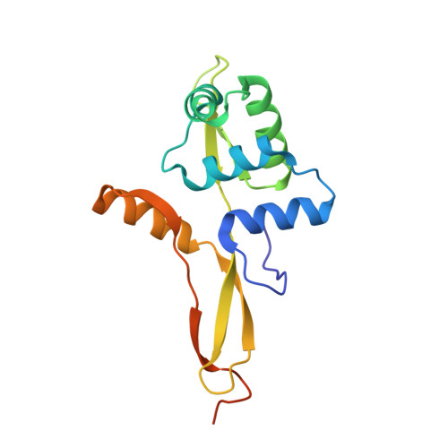
Group A streptococcus (GAS, Streptococcus pyogenes) is a strict human pathogen that causes severe, invasive diseases. GAS does not produce catalase, but has an ability to resist killing by reactive oxygen species (ROS) through novel mechanisms. The peroxide response regulator (PerR), a member of ferric uptake regulator (Fur) family, plays a key role for GAS to cope with oxidative stress by regulating the expression of multiple genes. Our previous studies have found that expression of an iron-binding protein, Dpr, is under the direct control of PerR. To elucidate the molecular interactions of PerR with its cognate promoter, we have carried out structural studies on PerR and PerR-DNA complex. By combining crystallography and small-angle X-ray scattering (SAXS), we confirmed that the determined PerR crystal structure reflects its conformation in solution. Through mutagenesis and biochemical analysis, we have identified DNA-binding residues suggesting that PerR binds to the dpr promoter at the per box through a winged-helix motif. Furthermore, we have performed SAXS analysis and resolved the molecular architecture of PerR-DNA complex, in which two 30 bp DNA fragments wrap around two PerR homodimers by interacting with the adjacent positively-charged winged-helix motifs. Overall, we provide structural insights into molecular recognition of DNA by PerR and define the hollow structural arrangement of PerR-30bpDNA complex, which displays a unique topology distinct from currently proposed DNA-binding models for Fur family regulators.
Organizational Affiliation:
Department of Microbiology and Immunology, College of Medicine, National Cheng Kung University, Tainan, Taiwan.