


4LEO
Crystal structure of anti-HER3 Fab RG7116 in complex with the extracellular domains of human Her3 (ERBB3)
- PDB DOI: https://doi.org/10.2210/pdb4LEO/pdb
- Classification: Transferase/Immune System
- Organism(s): Mus musculus, Homo sapiens
- Expression System: Cricetulus griseus
- Mutation(s): No
- Deposited: 2013-06-26 Released: 2013-07-10
Experimental Data Snapshot
- Method: X-RAY DIFFRACTION
- Resolution: 2.64 Å
- R-Value Free: 0.258
- R-Value Work: 0.228
- R-Value Observed: 0.230

This is version 2.0 of the entry. See complete history.

Macromolecules
Find similar proteins by:
(by identity cutoff) | 3D Structure
Entity ID: 1 | |||||
|---|---|---|---|---|---|
| Molecule | Chains | Sequence Length | Organism | Details | Image |
| RG7116 Fab heavy chain | 227 | Mus musculus | Mutation(s): 0 | 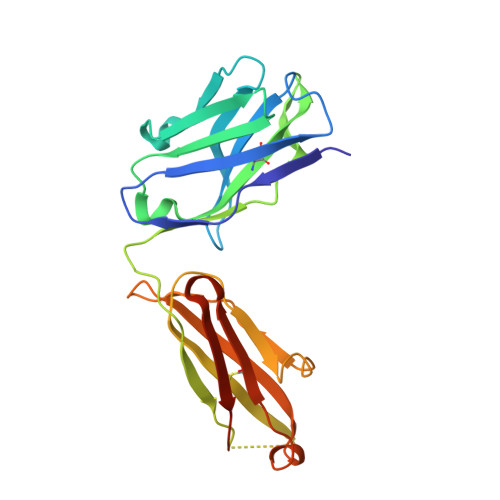 | |
Entity Groups | |||||
| Sequence Clusters | 30% Identity50% Identity70% Identity90% Identity95% Identity100% Identity | ||||
Sequence AnnotationsExpand | |||||
| |||||
Find similar proteins by:
(by identity cutoff) | 3D Structure
Entity ID: 2 | |||||
|---|---|---|---|---|---|
| Molecule | Chains | Sequence Length | Organism | Details | Image |
| RG7116 Fab light chain | 220 | Mus musculus | Mutation(s): 0 | 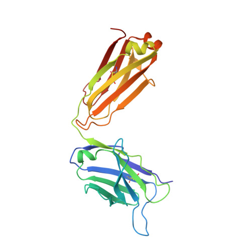 | |
Entity Groups | |||||
| Sequence Clusters | 30% Identity50% Identity70% Identity90% Identity95% Identity100% Identity | ||||
Sequence AnnotationsExpand | |||||
| |||||
Find similar proteins by:
(by identity cutoff) | 3D Structure
Entity ID: 3 | |||||
|---|---|---|---|---|---|
| Molecule | Chains | Sequence Length | Organism | Details | Image |
| Receptor tyrosine-protein kinase erbB-3 | 621 | Homo sapiens | Mutation(s): 0 Gene Names: ERBB3, HER3 EC: 2.7.10.1 | 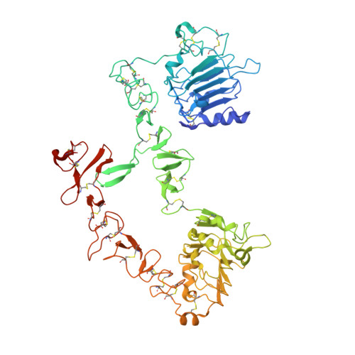 | |
UniProt & NIH Common Fund Data Resources | |||||
Find proteins for P21860 (Homo sapiens) Explore P21860 Go to UniProtKB: P21860 | |||||
PHAROS: P21860 GTEx: ENSG00000065361 | |||||
Entity Groups | |||||
| Sequence Clusters | 30% Identity50% Identity70% Identity90% Identity95% Identity100% Identity | ||||
| UniProt Group | P21860 | ||||
Sequence AnnotationsExpand | |||||
| |||||
Oligosaccharides

Small Molecules
| Ligands 1 Unique | |||||
|---|---|---|---|---|---|
| ID | Chains | Name / Formula / InChI Key | 2D Diagram | 3D Interactions | |
| NAG Query on NAG | E [auth C], F [auth C], G [auth C], H [auth C], I [auth C] | 2-acetamido-2-deoxy-beta-D-glucopyranose C8 H15 N O6 OVRNDRQMDRJTHS-FMDGEEDCSA-N |  | ||
Experimental Data & Validation
Experimental Data
- Method: X-RAY DIFFRACTION
- Resolution: 2.64 Å
- R-Value Free: 0.258
- R-Value Work: 0.228
- R-Value Observed: 0.230
- Space Group: P 1
Unit Cell:
| Length ( Å ) | Angle ( ˚ ) |
|---|---|
| a = 54.458 | α = 97.64 |
| b = 59.359 | β = 106.85 |
| c = 103.661 | γ = 96.83 |
| Software Name | Purpose |
|---|---|
| PHASER | phasing |
| PHENIX | refinement |
| XDS | data reduction |
| XDS | data scaling |
Entry History
Deposition Data
- Released Date: 2013-07-10 Deposition Author(s): Schiller, C.B., Hopfner, K.P.
Revision History (Full details and data files)
- Version 1.0: 2013-07-10
Type: Initial release - Version 1.1: 2013-08-28
Changes: Database references - Version 1.2: 2017-11-15
Changes: Advisory, Refinement description - Version 1.3: 2018-01-24
Changes: Structure summary - Version 2.0: 2020-07-29
Type: Remediation
Reason: Carbohydrate remediation
Changes: Atomic model, Data collection, Database references, Derived calculations, Structure summary
