4L40

Structure of the P450 OleT with a C20 fatty acid substrate bound


Experimental Data Snapshot

  • Method: X-RAY DIFFRACTION
  • Resolution: 2.50 Å
  • R-Value Free: 0.269 
  • R-Value Work: 0.195 
  • R-Value Observed: 0.199 

wwPDB Validation   3D Report Full Report


Ligand Structure Quality Assessment 


This is version 1.3 of the entry. See complete history


Literature

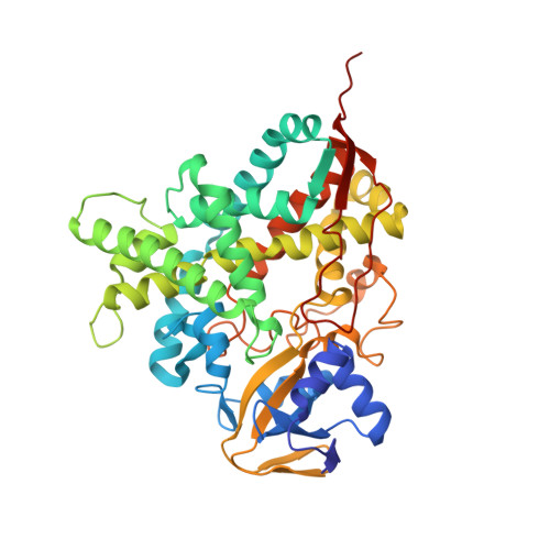
Structure and Biochemical Properties of the Alkene Producing Cytochrome P450 OleTJE (CYP152L1) from the Jeotgalicoccus sp. 8456 Bacterium.

Belcher, J.McLean, K.J.Matthews, S.Woodward, L.S.Fisher, K.Rigby, S.E.Nelson, D.R.Potts, D.Baynham, M.T.Parker, D.A.Leys, D.Munro, A.W.

(2014) J Biol Chem 289: 6535-6550

  • DOI: https://doi.org/10.1074/jbc.M113.527325
  • Primary Citation of Related Structures:  
    4L40, 4L54

  • PubMed Abstract: 

    The production of hydrocarbons in nature has been documented for only a limited set of organisms, with many of the molecular components underpinning these processes only recently identified. There is an obvious scope for application of these catalysts and engineered variants thereof in the future production of biofuels. Here we present biochemical characterization and crystal structures of a cytochrome P450 fatty acid peroxygenase: the terminal alkene forming OleTJE (CYP152L1) from Jeotgalicoccus sp. 8456. OleTJE is stabilized at high ionic strength, but aggregation and precipitation of OleTJE in low salt buffer can be turned to advantage for purification, because resolubilized OleTJE is fully active and extensively dissociated from lipids. OleTJE binds avidly to a range of long chain fatty acids, and structures of both ligand-free and arachidic acid-bound OleTJE reveal that the P450 active site is preformed for fatty acid binding. OleTJE heme iron has an unusually positive redox potential (-103 mV versus normal hydrogen electrode), which is not significantly affected by substrate binding, despite extensive conversion of the heme iron to a high spin ferric state. Terminal alkenes are produced from a range of saturated fatty acids (C12-C20), and stopped-flow spectroscopy indicates a rapid reaction between peroxide and fatty acid-bound OleTJE (167 s(-1) at 200 μm H2O2). Surprisingly, the active site is highly similar in structure to the related P450BSβ, which catalyzes hydroxylation of fatty acids as opposed to decarboxylation. Our data provide new insights into structural and mechanistic properties of a robust P450 with potential industrial applications.


  • Organizational Affiliation

    Manchester Institute of Biotechnology, Faculty of Life Sciences, University of Manchester, Manchester M1 7DN, United Kingdom.


Macromolecules
Find similar proteins by:  (by identity cutoff)  |  3D Structure
Entity ID: 1
MoleculeChains Sequence LengthOrganismDetailsImage
Terminal olefin-forming fatty acid decarboxylase422Jeotgalicoccus sp. ATCC 8456Mutation(s): 0 
UniProt
Find proteins for E9NSU2 (Jeotgalicoccus sp. ATCC 8456)
Explore E9NSU2 
Go to UniProtKB:  E9NSU2
Entity Groups  
Sequence Clusters30% Identity50% Identity70% Identity90% Identity95% Identity100% Identity
UniProt GroupE9NSU2
Sequence Annotations
Expand
  • Reference Sequence
Small Molecules
Ligands 2 Unique
IDChains Name / Formula / InChI Key2D Diagram3D Interactions
HEM
Query on HEM

Download Ideal Coordinates CCD File 
B [auth A]PROTOPORPHYRIN IX CONTAINING FE
C34 H32 Fe N4 O4
KABFMIBPWCXCRK-RGGAHWMASA-L
DCR
Query on DCR

Download Ideal Coordinates CCD File 
C [auth A]icosanoic acid
C20 H40 O2
VKOBVWXKNCXXDE-UHFFFAOYSA-N
Binding Affinity Annotations 
IDSourceBinding Affinity
DCR Binding MOAD:  4L40 Kd: 290 (nM) from 1 assay(s)
Experimental Data & Validation

Experimental Data

  • Method: X-RAY DIFFRACTION
  • Resolution: 2.50 Å
  • R-Value Free: 0.269 
  • R-Value Work: 0.195 
  • R-Value Observed: 0.199 
  • Space Group: P 43 21 2
Unit Cell:
Length ( Å )Angle ( ˚ )
a = 59.63α = 90
b = 59.63β = 90
c = 244.17γ = 90
Software Package:
Software NamePurpose
ADSCdata collection
PHASERphasing
REFMACrefinement
XDSdata reduction
XDSdata scaling

Structure Validation

View Full Validation Report


Ligand Structure Quality Assessment 


Entry History 

Deposition Data

  • Released Date: 2013-11-27 
  • Deposition Author(s): Leys, D.

Revision History  (Full details and data files)

  • Version 1.0: 2013-11-27
    Type: Initial release
  • Version 1.1: 2014-03-05
    Changes: Database references
  • Version 1.2: 2014-05-14
    Changes: Database references
  • Version 1.3: 2023-09-20
    Changes: Data collection, Database references, Derived calculations, Refinement description