4KRI

Haemonchus contortus Phospholethanolamine N-methyltransferase 2 in complex with phosphomonomethylethanolamine and S-adenosylhomocysteine


Experimental Data Snapshot

  • Method: X-RAY DIFFRACTION
  • Resolution: 1.72 Å
  • R-Value Free: 0.210 
  • R-Value Work: 0.174 
  • R-Value Observed: 0.176 

wwPDB Validation   3D Report Full Report


Ligand Structure Quality Assessment 


This is version 1.2 of the entry. See complete history


Literature

Evolution of structure and mechanistic divergence in di-domain methyltransferases from nematode phosphocholine biosynthesis.

Lee, S.G.Jez, J.M.

(2013) Structure 21: 1778-1787

  • DOI: https://doi.org/10.1016/j.str.2013.07.023
  • Primary Citation of Related Structures:  
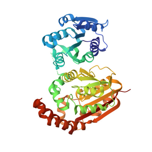
    4KRH, 4KRI

  • PubMed Abstract: 

    The phosphobase methylation pathway is the major route for supplying phosphocholine to phospholipid biosynthesis in plants, nematodes, and Plasmodium. In this pathway, phosphoethanolamine N-methyltransferase (PMT) catalyzes the sequential methylation of phosphoethanolamine to phosphocholine. In the PMT, one domain (MT1) catalyzes methylation of phosphoethanolamine to phosphomonomethylethanolamine and a second domain (MT2) completes the synthesis of phosphocholine. The X-ray crystal structures of the di-domain PMT from the parasitic nematode Haemonchus contortus (HcPMT1 and HcPMT2) reveal that the catalytic domains of these proteins are structurally distinct and allow for selective methylation of phosphobase substrates using different active site architectures. These structures also reveal changes leading to loss of function in the vestigial domains of the nematode PMT. Divergence of function in the two nematode PMTs provides two distinct antiparasitic inhibitor targets within the same essential metabolic pathway. The PMTs from nematodes, plants, and Plasmodium also highlight adaptable metabolic modularity in evolutionarily diverse organisms.


  • Organizational Affiliation

    Department of Biology, Washington University in St. Louis, One Brookings Drive, Campus Box 1137, St. Louis, MO 63130, USA.


Macromolecules
Find similar proteins by:  (by identity cutoff)  |  3D Structure
Entity ID: 1
MoleculeChains Sequence LengthOrganismDetailsImage
Phospholethanolamine N-methyltransferase 2
A, B, C
433Haemonchus contortusMutation(s): 0 
UniProt
Find proteins for U5HK48 (Haemonchus contortus)
Explore U5HK48 
Go to UniProtKB:  U5HK48
Entity Groups  
Sequence Clusters30% Identity50% Identity70% Identity90% Identity95% Identity100% Identity
UniProt GroupU5HK48
Sequence Annotations
Expand
  • Reference Sequence
Experimental Data & Validation

Experimental Data

  • Method: X-RAY DIFFRACTION
  • Resolution: 1.72 Å
  • R-Value Free: 0.210 
  • R-Value Work: 0.174 
  • R-Value Observed: 0.176 
  • Space Group: C 1 2 1
Unit Cell:
Length ( Å )Angle ( ˚ )
a = 135.987α = 90
b = 136.838β = 118.39
c = 90.867γ = 90
Software Package:
Software NamePurpose
HKL-3000data collection
PHASERphasing
PHENIXrefinement
HKL-3000data reduction
HKL-3000data scaling

Structure Validation

View Full Validation Report


Ligand Structure Quality Assessment 


Entry History 

Deposition Data

Revision History  (Full details and data files)

  • Version 1.0: 2013-09-25
    Type: Initial release
  • Version 1.1: 2013-10-30
    Changes: Database references
  • Version 1.2: 2024-02-28
    Changes: Data collection, Database references, Derived calculations