4KKD

The X-ray crystal structure of Mannose-binding lectin-associated serine proteinase-3 reveals the structural basis for enzyme inactivity associated with the 3MC syndrome


Experimental Data Snapshot

  • Method: X-RAY DIFFRACTION
  • Resolution: 2.60 Å
  • R-Value Free: 0.260 
  • R-Value Work: 0.194 
  • R-Value Observed: 0.196 

wwPDB Validation   3D Report Full Report


This is version 1.2 of the entry. See complete history


Literature

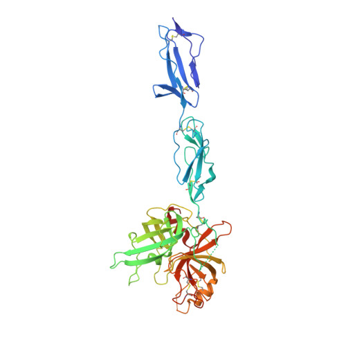
The X-ray Crystal Structure of Mannose-binding Lectin-associated Serine Proteinase-3 Reveals the Structural Basis for Enzyme Inactivity Associated with the Carnevale, Mingarelli, Malpuech, and Michels (3MC) Syndrome.

Yongqing, T.Wilmann, P.G.Reeve, S.B.Coetzer, T.H.Smith, A.I.Whisstock, J.C.Pike, R.N.Wijeyewickrema, L.C.

(2013) J Biol Chem 288: 22399-22407

  • DOI: https://doi.org/10.1074/jbc.M113.483875
  • Primary Citation of Related Structures:  
    4KKD

  • PubMed Abstract: 

    The mannose-binding lectin associated-protease-3 (MASP-3) is a member of the lectin pathway of the complement system, a key component of human innate and active immunity. Mutations in MASP-3 have recently been found to be associated with Carnevale, Mingarelli, Malpuech, and Michels (3MC) syndrome, a severe developmental disorder manifested by cleft palate, intellectual disability, and skeletal abnormalities. However, the molecular basis for MASP-3 function remains to be understood. Here we characterize the substrate specificity of MASP-3 by screening against a combinatorial peptide substrate library. Through this approach, we successfully identified a peptide substrate that was 20-fold more efficiently cleaved than any other identified to date. Furthermore, we demonstrated that mutant forms of the enzyme associated with 3MC syndrome were completely inactive against this substrate. To address the structural basis for this defect, we determined the 2.6-Å structure of the zymogen form of the G666E mutant of MASP-3. These data reveal that the mutation disrupts the active site and perturbs the position of the catalytic serine residue. Together, these insights into the function of MASP-3 reveal how a mutation in this enzyme causes it to be inactive and thus contribute to the 3MC syndrome.


  • Organizational Affiliation

    Department of Biochemistry & Molecular Biology, Monash University, Clayton, Victoria 3800, Australia.


Macromolecules
Find similar proteins by:  (by identity cutoff)  |  3D Structure
Entity ID: 1
MoleculeChains Sequence LengthOrganismDetailsImage
Mannan-binding lectin serine protease 1
A, B
433Homo sapiensMutation(s): 2 
Gene Names: CRARFCRARF1MASP1PRSS5
EC: 3.4.21
UniProt & NIH Common Fund Data Resources
Find proteins for P48740 (Homo sapiens)
Explore P48740 
Go to UniProtKB:  P48740
PHAROS:  P48740
GTEx:  ENSG00000127241 
Entity Groups  
Sequence Clusters30% Identity50% Identity70% Identity90% Identity95% Identity100% Identity
UniProt GroupP48740
Sequence Annotations
Expand
  • Reference Sequence
Small Molecules
Ligands 1 Unique
IDChains Name / Formula / InChI Key2D Diagram3D Interactions
IMD
Query on IMD

Download Ideal Coordinates CCD File 
C [auth B]IMIDAZOLE
C3 H5 N2
RAXXELZNTBOGNW-UHFFFAOYSA-O
Experimental Data & Validation

Experimental Data

  • Method: X-RAY DIFFRACTION
  • Resolution: 2.60 Å
  • R-Value Free: 0.260 
  • R-Value Work: 0.194 
  • R-Value Observed: 0.196 
  • Space Group: P 21 21 2
Unit Cell:
Length ( Å )Angle ( ˚ )
a = 72.84α = 90
b = 292.916β = 90
c = 43.589γ = 90
Software Package:
Software NamePurpose
ADSCdata collection
Auto-Rickshawphasing
PHENIXrefinement
XDSdata reduction
pointlessdata scaling

Structure Validation

View Full Validation Report


Entry History 

Deposition Data

Revision History  (Full details and data files)

  • Version 1.0: 2013-07-03
    Type: Initial release
  • Version 1.1: 2013-07-10
    Changes: Structure summary
  • Version 1.2: 2013-08-21
    Changes: Database references