The landscape of cytokinin binding by a plant nodulin.
Ruszkowski, M., Szpotkowski, K., Sikorski, M., Jaskolski, M.(2013) Acta Crystallogr D Biol Crystallogr 69: 2365-2380
- PubMed: 24311578
- DOI: https://doi.org/10.1107/S0907444913021975
- Primary Citation of Related Structures:
4GY9, 4JHG, 4JHH, 4JHI - PubMed Abstract:
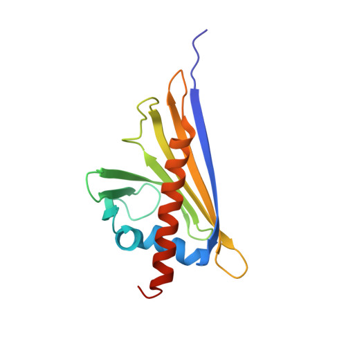
Nodulation is an extraordinary symbiotic interaction between leguminous plants and nitrogen-fixing bacteria (rhizobia) that assimilate atmospheric nitrogen (in root nodules) and convert it into compounds suitable for the plant host. A class of plant hormones called cytokinins are involved in the nodulation process. In the model legume Medicago truncatula, nodulin 13 (MtN13), which belongs to the pathogenesis-related proteins of class 10 (PR-10), is expressed in the outer cortex of the nodules. In general, PR-10 proteins are small and monomeric and have a characteristic fold with an internal hydrophobic cavity formed between a seven-stranded antiparallel β-sheet and a C-terminal α-helix. Previously, some PR-10 proteins not related to nodulation were found to bind cytokinins such as trans-zeatin. Here, four crystal structures of the MtN13 protein are reported in complexes with several cytokinins, namely trans-zeatin, N6-isopentenyladenine, kinetin and N6-benzyladenine. All four phytohormones are bound in the hydrophobic cavity in the same manner and have excellent definition in the electron-density maps. The binding of the cytokinins appears to be strong and specific and is reinforced by several hydrogen bonds. Although the binding stoichiometry is 1:1, the complex is actually dimeric, with a cytokinin molecule bound in each subunit. The ligand-binding site in each cavity is formed with the participation of a loop element from the other subunit, which plugs the only entrance to the cavity. Interestingly, a homodimer of MtN13 is also formed in solution, as confirmed by small-angle X-ray scattering (SAXS).
Organizational Affiliation:
Center for Biocrystallographic Research, Institute of Bioorganic Chemistry, Polish Academy of Sciences, Poznan, Poland.