4JCV

Crystal structure of the RecOR complex in an open conformation


Experimental Data Snapshot

  • Method: X-RAY DIFFRACTION
  • Resolution: 3.34 Å
  • R-Value Free: 0.278 
  • R-Value Work: 0.240 
  • R-Value Observed: 0.242 

wwPDB Validation   3D Report Full Report


This is version 1.2 of the entry. See complete history


Literature

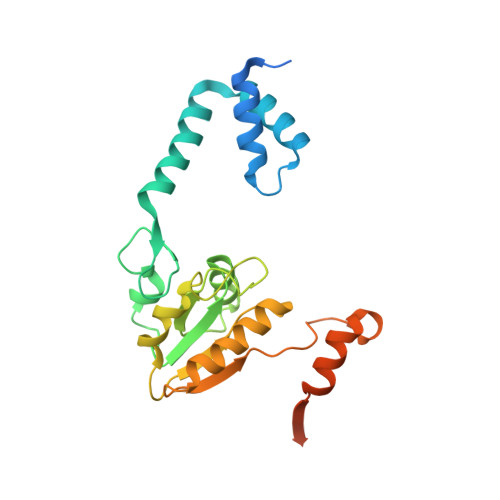
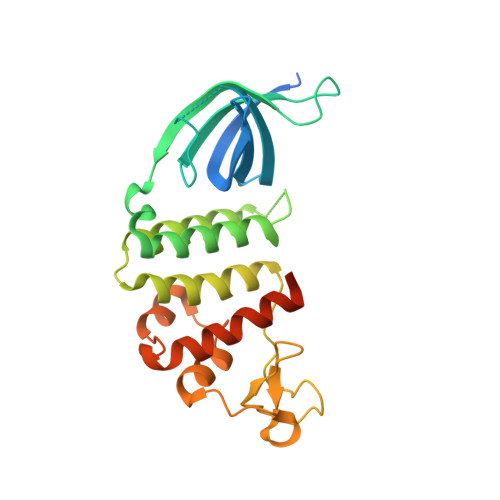
An 'open' structure of the RecOR complex supports ssDNA binding within the core of the complex.

Radzimanowski, J.Dehez, F.Round, A.Bidon-Chanal, A.McSweeney, S.Timmins, J.

(2013) Nucleic Acids Res 41: 7972-7986

  • DOI: https://doi.org/10.1093/nar/gkt572
  • Primary Citation of Related Structures:  
    4JCV

  • PubMed Abstract: 

    Efficient DNA repair is critical for cell survival and the maintenance of genome integrity. The homologous recombination pathway is responsible for the repair of DNA double-strand breaks within cells. Initiation of this pathway in bacteria can be carried out by either the RecBCD or the RecFOR proteins. An important regulatory player within the RecFOR pathway is the RecOR complex that facilitates RecA loading onto DNA. Here we report new data regarding the assembly of Deinococcus radiodurans RecOR and its interaction with DNA, providing novel mechanistic insight into the mode of action of RecOR in homologous recombination. We present a higher resolution crystal structure of RecOR in an 'open' conformation in which the tetrameric RecR ring flanked by two RecO molecules is accessible for DNA binding. We show using small-angle neutron scattering and mutagenesis studies that DNA binding does indeed occur within the RecR ring. Binding of single-stranded DNA occurs without any major conformational changes of the RecOR complex while structural rearrangements are observed on double-stranded DNA binding. Finally, our molecular dynamics simulations, supported by our biochemical data, provide a detailed picture of the DNA binding motif of RecOR and reveal that single-stranded DNA is sandwiched between the two facing oligonucleotide binding domains of RecO within the RecR ring.


  • Organizational Affiliation

    Structural Biology Group, European Synchrotron Radiation Facility, 6 rue Jules Horowitz, 38043 Grenoble cedex 9, France, Université de Lorraine, BP239, 54506 Vandoeuvre-lès-Nancy Cedex, France, CNRS, UMR N°7565, 54506 Vandoeuvre-les-Nancy, France, European Molecular Biology Laboratory, Grenoble Outstation, 6 rue Jules Horowitz, 38042 Grenoble, France, Unit for Virus Host-Cell Interactions, Univ. Grenoble Alpes-EMBL-CNRS, 6 rue Jules Horowitz, 38042 Grenoble, France and Institut de Biologie Structurale, CNRS/CEA/Université de Grenoble, 41 rue Jules Horowitz, 38027 Grenoble cedex 1, France.


Macromolecules
Find similar proteins by:  (by identity cutoff)  |  3D Structure
Entity ID: 1
MoleculeChains Sequence LengthOrganismDetailsImage
Recombination protein RecR
A, B, C, D
241Deinococcus radiodurans R1 = ATCC 13939 = DSM 20539Mutation(s): 2 
Gene Names: recRDR_0198
UniProt
Find proteins for Q9ZNA2 (Deinococcus radiodurans (strain ATCC 13939 / DSM 20539 / JCM 16871 / CCUG 27074 / LMG 4051 / NBRC 15346 / NCIMB 9279 / VKM B-1422 / R1))
Explore Q9ZNA2 
Go to UniProtKB:  Q9ZNA2
Entity Groups  
Sequence Clusters30% Identity50% Identity70% Identity90% Identity95% Identity100% Identity
UniProt GroupQ9ZNA2
Sequence Annotations
Expand
  • Reference Sequence
Find similar proteins by:  (by identity cutoff)  |  3D Structure
Entity ID: 2
MoleculeChains Sequence LengthOrganismDetailsImage
DNA repair protein RecO
E, F
265Deinococcus radiodurans R1 = ATCC 13939 = DSM 20539Mutation(s): 0 
Gene Names: recODR_0819
UniProt
Find proteins for Q9RW50 (Deinococcus radiodurans (strain ATCC 13939 / DSM 20539 / JCM 16871 / CCUG 27074 / LMG 4051 / NBRC 15346 / NCIMB 9279 / VKM B-1422 / R1))
Explore Q9RW50 
Go to UniProtKB:  Q9RW50
Entity Groups  
Sequence Clusters30% Identity50% Identity70% Identity90% Identity95% Identity100% Identity
UniProt GroupQ9RW50
Sequence Annotations
Expand
  • Reference Sequence
Experimental Data & Validation

Experimental Data

  • Method: X-RAY DIFFRACTION
  • Resolution: 3.34 Å
  • R-Value Free: 0.278 
  • R-Value Work: 0.240 
  • R-Value Observed: 0.242 
  • Space Group: P 1
Unit Cell:
Length ( Å )Angle ( ˚ )
a = 63.13α = 103.6
b = 93.11β = 110.42
c = 92.35γ = 106.22
Software Package:
Software NamePurpose
ADSCdata collection
PHASERphasing
REFMACrefinement
MOSFLMdata reduction
SCALAdata scaling

Structure Validation

View Full Validation Report


Entry History 

Deposition Data

Revision History  (Full details and data files)

  • Version 1.0: 2013-08-07
    Type: Initial release
  • Version 1.1: 2014-10-01
    Changes: Database references
  • Version 1.2: 2023-11-08
    Changes: Data collection, Database references, Derived calculations, Refinement description