4JC6

Mercury activation of the plant aquaporin SoPIP2;1 - structural and functional characterization


Experimental Data Snapshot

  • Method: X-RAY DIFFRACTION
  • Resolution: 2.15 Å
  • R-Value Free: 0.227 
  • R-Value Work: 0.197 
  • R-Value Observed: 0.198 

wwPDB Validation   3D Report Full Report


Ligand Structure Quality Assessment 


This is version 1.4 of the entry. See complete history


Literature

Mercury increases water permeability of a plant aquaporin through a non-cysteine-related mechanism.

Frick, A.Jarva, M.Ekvall, M.Uzdavinys, P.Nyblom, M.Tornroth-Horsefield, S.

(2013) Biochem J 454: 491-499

  • DOI: https://doi.org/10.1042/BJ20130377
  • Primary Citation of Related Structures:  
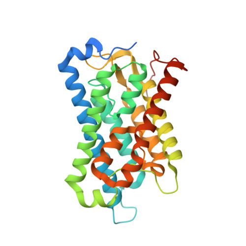
    4JC6

  • PubMed Abstract: 

    Water transport across cellular membranes is mediated by a family of membrane proteins known as AQPs (aquaporins). AQPs were first discovered on the basis of their ability to be inhibited by mercurial compounds, an experiment which has followed the AQP field ever since. Although mercury inhibition is most common, many AQPs are mercury insensitive. In plants, regulation of AQPs is important in order to cope with environmental changes. Plant plasma membrane AQPs are known to be gated by phosphorylation, pH and Ca²⁺. We have previously solved the structure of the spinach AQP SoPIP2;1 (Spinacia oleracea plasma membrane intrinsic protein 2;1) in closed and open conformations and proposed a mechanism for how this gating can be achieved. To study the effect of mercury on SoPIP2;1 we solved the structure of the SoPIP2;1-mercury complex and characterized the water transport ability using proteoliposomes. The structure revealed mercury binding to three out of four cysteine residues. In contrast to what is normally seen for AQPs, mercury increased the water transport rate of SoPIP2;1, an effect which could not be attributed to any of the cysteine residues. This indicates that other factors might influence the effect of mercury on SoPIP2;1, one of which could be the properties of the lipid bilayer.


  • Organizational Affiliation

    Department of Chemistry and Molecular Biology, University of Gothenburg, PO Box 462, SE-405 30 Gothenburg, Sweden.


Macromolecules
Find similar proteins by:  (by identity cutoff)  |  3D Structure
Entity ID: 1
MoleculeChains Sequence LengthOrganismDetailsImage
Aquaporin281Spinacia oleraceaMutation(s): 0 
Membrane Entity: Yes 
UniProt
Find proteins for Q41372 (Spinacia oleracea)
Explore Q41372 
Go to UniProtKB:  Q41372
Entity Groups  
Sequence Clusters30% Identity50% Identity70% Identity90% Identity95% Identity100% Identity
UniProt GroupQ41372
Sequence Annotations
Expand
  • Reference Sequence
Small Molecules
Ligands 3 Unique
IDChains Name / Formula / InChI Key2D Diagram3D Interactions
BOG
Query on BOG

Download Ideal Coordinates CCD File 
AA [auth C]
EB [auth L]
FB [auth L]
GA [auth D]
HA [auth D]
AA [auth C],
EB [auth L],
FB [auth L],
GA [auth D],
HA [auth D],
I [auth A],
J [auth A],
K [auth A],
LB [auth N],
MB [auth N],
NA [auth H],
OA [auth H],
WA [auth J]
octyl beta-D-glucopyranoside
C14 H28 O6
HEGSGKPQLMEBJL-RKQHYHRCSA-N
HG
Query on HG

Download Ideal Coordinates CCD File 
AB [auth J]
BB [auth J]
CB [auth J]
DA [auth C]
DB [auth J]
AB [auth J],
BB [auth J],
CB [auth J],
DA [auth C],
DB [auth J],
EA [auth C],
FA [auth C],
IB [auth L],
JB [auth L],
KA [auth D],
KB [auth L],
LA [auth D],
MA [auth D],
O [auth A],
P [auth A],
PB [auth N],
Q [auth A],
QB [auth N],
R [auth A],
RA [auth H],
RB [auth N],
S [auth A],
SA [auth H],
TA [auth H],
UA [auth H],
V [auth B],
VA [auth H],
W [auth B],
X [auth B],
Y [auth B],
Z [auth B],
ZA [auth J]
MERCURY (II) ION
Hg
BQPIGGFYSBELGY-UHFFFAOYSA-N
CD
Query on CD

Download Ideal Coordinates CCD File 
BA [auth C]
CA [auth C]
GB [auth L]
HB [auth L]
IA [auth D]
BA [auth C],
CA [auth C],
GB [auth L],
HB [auth L],
IA [auth D],
JA [auth D],
L [auth A],
M [auth A],
N [auth A],
NB [auth N],
OB [auth N],
PA [auth H],
QA [auth H],
T [auth B],
U [auth B],
XA [auth J],
YA [auth J]
CADMIUM ION
Cd
WLZRMCYVCSSEQC-UHFFFAOYSA-N
Experimental Data & Validation

Experimental Data

  • Method: X-RAY DIFFRACTION
  • Resolution: 2.15 Å
  • R-Value Free: 0.227 
  • R-Value Work: 0.197 
  • R-Value Observed: 0.198 
  • Space Group: P 21 21 21
Unit Cell:
Length ( Å )Angle ( ˚ )
a = 123.546α = 90
b = 141.818β = 90
c = 186.515γ = 90
Software Package:
Software NamePurpose
SCALEPACKdata scaling
MOLREPphasing
REFMACrefinement
PDB_EXTRACTdata extraction
DENZOdata reduction

Structure Validation

View Full Validation Report


Ligand Structure Quality Assessment 


Entry History 

Deposition Data

Revision History  (Full details and data files)

  • Version 1.0: 2013-09-11
    Type: Initial release
  • Version 1.1: 2015-02-04
    Changes: Structure summary
  • Version 1.2: 2017-11-15
    Changes: Refinement description
  • Version 1.3: 2020-07-29
    Type: Remediation
    Reason: Carbohydrate remediation
    Changes: Data collection, Derived calculations, Structure summary
  • Version 1.4: 2023-09-20
    Changes: Data collection, Database references, Refinement description, Structure summary