


Crystal structure of 16S ribosomal RNA methyltransferase RsmE
Eswaramoorthy, S., Almo, S.C., Swaminathan, S.To be published.
Experimental Data Snapshot
wwPDB Validation 3D Report Full Report

Entity ID: 1 | |||||
|---|---|---|---|---|---|
| Molecule | Chains | Sequence Length | Organism | Details | Image |
| Ribosomal RNA small subunit methyltransferase E | 267 | Sinorhizobium meliloti 1021 | Mutation(s): 0 Gene Names: R00771, SMc00826 EC: 2.1.1.193 | 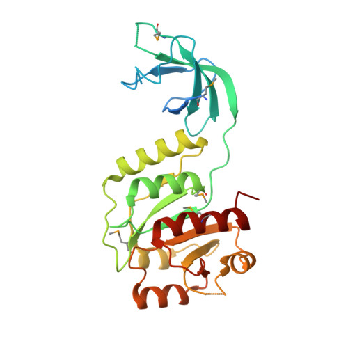 | |
UniProt | |||||
Find proteins for Q92RS8 (Rhizobium meliloti (strain 1021)) Explore Q92RS8 Go to UniProtKB: Q92RS8 | |||||
Entity Groups | |||||
| Sequence Clusters | 30% Identity50% Identity70% Identity90% Identity95% Identity100% Identity | ||||
| UniProt Group | Q92RS8 | ||||
Sequence AnnotationsExpand | |||||
| |||||
| Modified Residues 1 Unique | |||||
|---|---|---|---|---|---|
| ID | Chains | Type | Formula | 2D Diagram | Parent |
| MSE Query on MSE | A, B | L-PEPTIDE LINKING | C5 H11 N O2 Se |  | MET |
| Length ( Å ) | Angle ( ˚ ) |
|---|---|
| a = 96.085 | α = 90 |
| b = 152.082 | β = 90 |
| c = 38.995 | γ = 90 |
| Software Name | Purpose |
|---|---|
| CBASS | data collection |
| SHELXS | phasing |
| REFMAC | refinement |
| HKL-2000 | data reduction |
| HKL-2000 | data scaling |
RCSB PDB (citation) is hosted by
RCSB PDB is a member of the

