4IYN

Structure of mitochondrial Hsp90 (TRAP1) with ADP-ALF4-


Experimental Data Snapshot

  • Method: X-RAY DIFFRACTION
  • Resolution: 2.31 Å
  • R-Value Free: 0.231 
  • R-Value Work: 0.191 
  • R-Value Observed: 0.192 

wwPDB Validation   3D Report Full Report


Ligand Structure Quality Assessment 


This is version 1.2 of the entry. See complete history


Literature

Structural asymmetry in the closed state of mitochondrial Hsp90 (TRAP1) supports a two-step ATP hydrolysis mechanism.

Lavery, L.A.Partridge, J.R.Ramelot, T.A.Elnatan, D.Kennedy, M.A.Agard, D.A.

(2014) Mol Cell 53: 330-343

  • DOI: https://doi.org/10.1016/j.molcel.2013.12.023
  • Primary Citation of Related Structures:  
    4IPE, 4IVG, 4IYN, 4J0B

  • PubMed Abstract: 

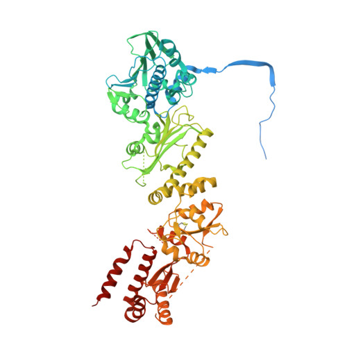
    While structural symmetry is a prevailing feature of homo-oligomeric proteins, asymmetry provides unique mechanistic opportunities. We present the crystal structure of full-length TRAP1, the mitochondrial Hsp90 molecular chaperone, in a catalytically active closed state. The TRAP1 homodimer adopts a distinct, asymmetric conformation, where one protomer is reconfigured via a helix swap at the middle:C-terminal domain (MD:CTD) interface. This interface plays a critical role in client binding. Solution methods validate the asymmetry and show extension to Hsp90 homologs. Point mutations that disrupt unique contacts at each MD:CTD interface reduce catalytic activity and substrate binding and demonstrate that each protomer needs access to both conformations. Crystallographic data on a dimeric NTD:MD fragment suggests that asymmetry arises from strain induced by simultaneous NTD and CTD dimerization. The observed asymmetry provides the potential for an additional step in the ATPase cycle, allowing sequential ATP hydrolysis steps to drive both client remodeling and client release.


  • Organizational Affiliation

    Howard Hughes Medical Institute and the Department of Biochemistry and Biophysics, University of California, San Francisco, San Francisco, CA 94158, USA.


Macromolecules
Find similar proteins by:  (by identity cutoff)  |  3D Structure
Entity ID: 1
MoleculeChains Sequence LengthOrganismDetailsImage
TNF receptor-associated protein 1
A, B
719Danio rerioMutation(s): 0 
Gene Names: trap1
UniProt
Find proteins for A8WFV1 (Danio rerio)
Explore A8WFV1 
Go to UniProtKB:  A8WFV1
Entity Groups  
Sequence Clusters30% Identity50% Identity70% Identity90% Identity95% Identity100% Identity
UniProt GroupA8WFV1
Sequence Annotations
Expand
  • Reference Sequence
Small Molecules
Ligands 4 Unique
IDChains Name / Formula / InChI Key2D Diagram3D Interactions
ADP
Query on ADP

Download Ideal Coordinates CCD File 
D [auth A],
J [auth B]
ADENOSINE-5'-DIPHOSPHATE
C10 H15 N5 O10 P2
XTWYTFMLZFPYCI-KQYNXXCUSA-N
ALF
Query on ALF

Download Ideal Coordinates CCD File 
E [auth A],
K [auth B]
TETRAFLUOROALUMINATE ION
Al F4
UYOMQIYKOOHAMK-UHFFFAOYSA-J
CO
Query on CO

Download Ideal Coordinates CCD File 
C [auth A],
G [auth A],
H [auth B],
I [auth B]
COBALT (II) ION
Co
XLJKHNWPARRRJB-UHFFFAOYSA-N
MG
Query on MG

Download Ideal Coordinates CCD File 
F [auth A],
L [auth B]
MAGNESIUM ION
Mg
JLVVSXFLKOJNIY-UHFFFAOYSA-N
Experimental Data & Validation

Experimental Data

  • Method: X-RAY DIFFRACTION
  • Resolution: 2.31 Å
  • R-Value Free: 0.231 
  • R-Value Work: 0.191 
  • R-Value Observed: 0.192 
  • Space Group: C 1 2 1
Unit Cell:
Length ( Å )Angle ( ˚ )
a = 178.896α = 90
b = 96.597β = 134.26
c = 125.901γ = 90
Software Package:
Software NamePurpose
Blu-Icedata collection
SOLVEphasing
PHENIXrefinement
HKL-2000data reduction
HKL-2000data scaling

Structure Validation

View Full Validation Report


Ligand Structure Quality Assessment 


Entry History 

Deposition Data

Revision History  (Full details and data files)

  • Version 1.0: 2014-01-22
    Type: Initial release
  • Version 1.1: 2014-08-27
    Changes: Database references
  • Version 1.2: 2024-04-03
    Changes: Data collection, Database references, Derived calculations, Refinement description