Structural Insights into the UbiD Protein Family from the Crystal Structure of PA0254 from Pseudomonas aeruginosa.
Jacewicz, A., Izumi, A., Brunner, K., Schnell, R., Schneider, G.(2013) PLoS One 8: e63161-e63161
- PubMed: 23671667
- DOI: https://doi.org/10.1371/journal.pone.0063161
- Primary Citation of Related Structures:
4IP2, 4IWS - PubMed Abstract:
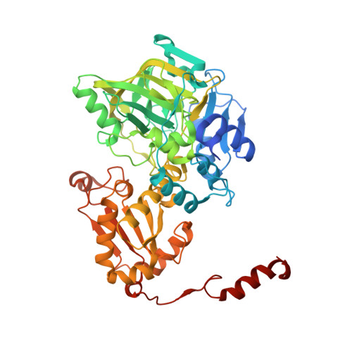
The 3-polyprenyl-4-hydroxybenzoate decarboxylase (UbiD) catalyzes the conversion of 3-polyprenyl-4-hydroxybenzoate to 2-polyprenylphenol in the biosynthesis of ubiquinone. Pseudomonas aeruginosa contains two genes (PA0254 and PA5237) that are related in sequence to putative UbiD enzymes. A bioinformatics analysis suggests that the UbiD sequence family can be divided into two subclasses, with PA5237 and PA0254 belonging to different branches of this family. The three-dimensional structure of PA0254 has been determined using single wavelength anomalous diffraction and molecular replacement in two different crystal forms to resolutions of 1.95 and 2.3 Å, respectively. The subunit of PA0254 consists of three domains, an N-terminal α/β domain, a split β-barrel with a similar fold of a family of flavin reductases and a C-terminal α/β domain with a topology characteristic for the UbiD protein family. The middle domain contains a metal binding site adjacent to a large open cleft that may represent the active site. The two protein ligands binding a magnesium ion, His188 and Glu229, invariant in the PA0254 subclass, are also conserved in a corresponding metal site found in one of the FMN binding proteins from the split β-barrel fold family. PA0254 forms, in contrast to the hexameric UbiD from E. coli and P. aeruginosa, a homo-dimer. Insertion of four residues in a loop region in the PA0254 type enzymes results in structural differences that are incompatible with hexamer assembly.
Organizational Affiliation:
Department of Medical Biochemistry and Biophysics, Karolinska Institutet, Stockholm, Sweden.