4IW3

Crystal structure of a Pseudomonas putida prolyl-4-hydroxylase (P4H) in complex with elongation factor Tu (EF-Tu)


Experimental Data Snapshot

  • Method: X-RAY DIFFRACTION
  • Resolution: 2.70 Å
  • R-Value Free: 0.218 
  • R-Value Work: 0.168 
  • R-Value Observed: 0.171 

wwPDB Validation   3D Report Full Report


Ligand Structure Quality Assessment 


This is version 1.5 of the entry. See complete history


Literature

Human oxygen sensing may have origins in prokaryotic elongation factor Tu prolyl-hydroxylation

Scotti, J.S.Leung, I.K.H.Ge, W.Bentley, M.A.Paps, J.Kramer, H.B.Lee, J.Aik, W.Choi, H.Paulsen, S.M.Bowman, L.A.H.Loik, N.D.Horita, S.Ho, C.H.Kershaw, N.J.Tang, C.M.Claridge, T.D.W.Preston, G.M.McDonough, M.A.Schofield, C.J.

(2014) Proc Natl Acad Sci U S A 111: 13331-13336

  • DOI: https://doi.org/10.1073/pnas.1409916111
  • Primary Citation of Related Structures:  
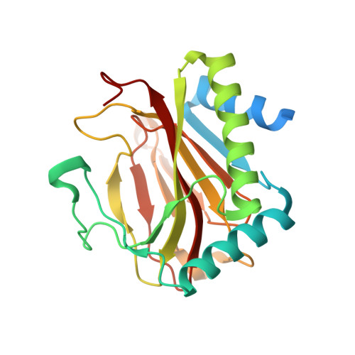
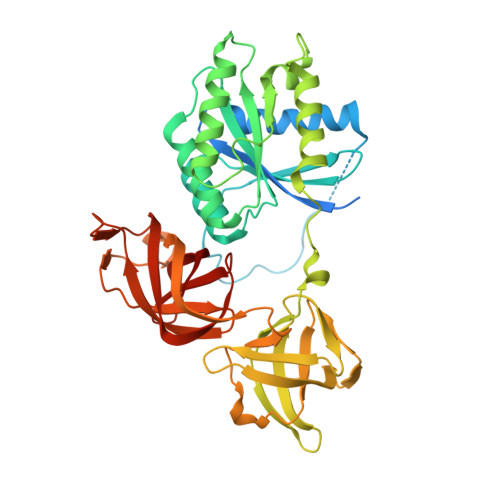
    4IW3, 4J0Q, 4J25

  • PubMed Abstract: 

    The roles of 2-oxoglutarate (2OG)-dependent prolyl-hydroxylases in eukaryotes include collagen stabilization, hypoxia sensing, and translational regulation. The hypoxia-inducible factor (HIF) sensing system is conserved in animals, but not in other organisms. However, bioinformatics imply that 2OG-dependent prolyl-hydroxylases (PHDs) homologous to those acting as sensing components for the HIF system in animals occur in prokaryotes. We report cellular, biochemical, and crystallographic analyses revealing that Pseudomonas prolyl-hydroxylase domain containing protein (PPHD) contain a 2OG oxygenase related in structure and function to the animal PHDs. A Pseudomonas aeruginosa PPHD knockout mutant displays impaired growth in the presence of iron chelators and increased production of the virulence factor pyocyanin. We identify elongation factor Tu (EF-Tu) as a PPHD substrate, which undergoes prolyl-4-hydroxylation on its switch I loop. A crystal structure of PPHD reveals striking similarity to human PHD2 and a Chlamydomonas reinhardtii prolyl-4-hydroxylase. A crystal structure of PPHD complexed with intact EF-Tu reveals that major conformational changes occur in both PPHD and EF-Tu, including a >20-Å movement of the EF-Tu switch I loop. Comparison of the PPHD structures with those of HIF and collagen PHDs reveals conservation in substrate recognition despite diverse biological roles and origins. The observed changes will be useful in designing new types of 2OG oxygenase inhibitors based on various conformational states, rather than active site iron chelators, which make up most reported 2OG oxygenase inhibitors. Structurally informed phylogenetic analyses suggest that the role of prolyl-hydroxylation in human hypoxia sensing has ancient origins.


  • Organizational Affiliation

    Chemistry Research Laboratory, Department of Chemistry, University of Oxford, Oxford OX1 3TA, United Kingdom;


Macromolecules
Find similar proteins by:  (by identity cutoff)  |  3D Structure
Entity ID: 1
MoleculeChains Sequence LengthOrganismDetailsImage
Putative uncharacterized proteinA,
D [auth J]
229Pseudomonas putida KT2440Mutation(s): 0 
Gene Names: PP_5159
UniProt
Find proteins for Q88CM1 (Pseudomonas putida (strain ATCC 47054 / DSM 6125 / CFBP 8728 / NCIMB 11950 / KT2440))
Explore Q88CM1 
Go to UniProtKB:  Q88CM1
Entity Groups  
Sequence Clusters30% Identity50% Identity70% Identity90% Identity95% Identity100% Identity
UniProt GroupQ88CM1
Sequence Annotations
Expand
  • Reference Sequence
Find similar proteins by:  (by identity cutoff)  |  3D Structure
Entity ID: 2
MoleculeChains Sequence LengthOrganismDetailsImage
Elongation factor Tu-AB,
C [auth K]
433Pseudomonas putida KT2440Mutation(s): 0 
Gene Names: PP_0440tuf-1tufA
UniProt
Find proteins for Q88QP8 (Pseudomonas putida (strain ATCC 47054 / DSM 6125 / CFBP 8728 / NCIMB 11950 / KT2440))
Explore Q88QP8 
Go to UniProtKB:  Q88QP8
Entity Groups  
Sequence Clusters30% Identity50% Identity70% Identity90% Identity95% Identity100% Identity
UniProt GroupQ88QP8
Sequence Annotations
Expand
  • Reference Sequence
Small Molecules
Ligands 4 Unique
IDChains Name / Formula / InChI Key2D Diagram3D Interactions
GDP
Query on GDP

Download Ideal Coordinates CCD File 
G [auth B],
J [auth K]
GUANOSINE-5'-DIPHOSPHATE
C10 H15 N5 O11 P2
QGWNDRXFNXRZMB-UUOKFMHZSA-N
OGA
Query on OGA

Download Ideal Coordinates CCD File 
F [auth A],
L [auth J]
N-OXALYLGLYCINE
C4 H5 N O5
BIMZLRFONYSTPT-UHFFFAOYSA-N
MN
Query on MN

Download Ideal Coordinates CCD File 
E [auth A],
K [auth J]
MANGANESE (II) ION
Mn
WAEMQWOKJMHJLA-UHFFFAOYSA-N
MG
Query on MG

Download Ideal Coordinates CCD File 
H [auth B],
I [auth K]
MAGNESIUM ION
Mg
JLVVSXFLKOJNIY-UHFFFAOYSA-N
Experimental Data & Validation

Experimental Data

  • Method: X-RAY DIFFRACTION
  • Resolution: 2.70 Å
  • R-Value Free: 0.218 
  • R-Value Work: 0.168 
  • R-Value Observed: 0.171 
  • Space Group: P 31 2 1
Unit Cell:
Length ( Å )Angle ( ˚ )
a = 200.704α = 90
b = 200.704β = 90
c = 74.833γ = 120
Software Package:
Software NamePurpose
XSCALEdata scaling
PHASERphasing
PHENIXrefinement
PDB_EXTRACTdata extraction
GDAdata collection

Structure Validation

View Full Validation Report


Ligand Structure Quality Assessment 


Entry History 

Deposition Data

Revision History  (Full details and data files)

  • Version 1.0: 2014-01-29
    Type: Initial release
  • Version 1.1: 2014-03-05
    Changes: Other
  • Version 1.2: 2014-09-17
    Changes: Database references
  • Version 1.3: 2014-10-01
    Changes: Database references
  • Version 1.4: 2019-11-20
    Changes: Data collection, Database references
  • Version 1.5: 2023-11-08
    Changes: Data collection, Database references, Derived calculations, Refinement description