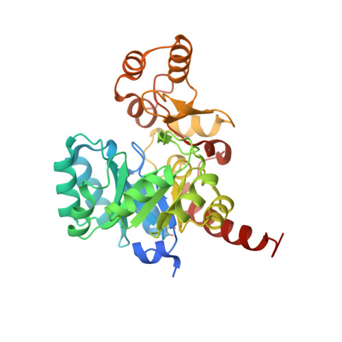
4IQL

Crystal Structure of Porphyromonas gingivalis Enoyl-ACP Reductase II (FabK) with cofactors NADPH and FMN


Experimental Data Snapshot

  • Method: X-RAY DIFFRACTION
  • Resolution: 1.94 Å
  • R-Value Free: 0.207 
  • R-Value Work: 0.161 
  • R-Value Observed: 0.163 

wwPDB Validation   3D Report Full Report


Ligand Structure Quality Assessment 


This is version 1.4 of the entry. See complete history


Literature

Structural characterization of Porphyromonas gingivalis enoyl-ACP reductase II (FabK).

Hevener, K.E.Santarsiero, B.D.Lee, H.Jones, J.A.Boci, T.Johnson, M.E.Mehboob, S.

(2018) Acta Crystallogr F Struct Biol Commun 74: 105-112

  • DOI: https://doi.org/10.1107/S2053230X18000262
  • Primary Citation of Related Structures:  
    4IQL

  • PubMed Abstract: 

    Enoyl-acyl carrier protein (ACP) reductase II (FabK) is a critical rate-limiting enzyme in the bacterial type II fatty-acid synthesis (FAS II) pathway. FAS II pathway enzymes are markedly disparate from their mammalian analogs in the FAS I pathway in both structure and mechanism. Enzymes involved in bacterial fatty-acid synthesis represent viable drug targets for Gram-negative pathogens, and historical precedent exists for targeting them in the treatment of diseases of the oral cavity. The Gram-negative organism Porphyromonas gingivalis represents a key causative agent of the costly and highly prevalent disease known as chronic periodontitis, and exclusively expresses FabK as its enoyl reductase enzyme in the FAS-II pathway. Together, these characteristics distinguish P. gingivalis FabK (PgFabK) as an attractive and novel narrow-spectrum antibacterial target candidate. PgFabK is a flavoenzyme that is dependent on FMN and NADPH as cofactors for the enzymatic reaction, which reduces the enoyl substrate via a ping-pong mechanism. Here, the structure of the PgFabK enzyme as determined using X-ray crystallography is reported to 1.9 Å resolution with endogenous FMN fully resolved and the NADPH cofactor partially resolved. PgFabK possesses a TIM-barrel motif, and all flexible loops are visible. The determined structure has allowed insight into the structural basis for the NADPH dependence observed in PgFabK and the role of a monovalent cation that has been observed in previous studies to be stringently required for FabK activity. The PgFabK structure and the insights gleaned from its analysis will facilitate structure-based drug-discovery efforts towards the prevention and treatment of P. gingivalis infection.


  • Organizational Affiliation

    Department of Pharmaceutical Sciences, University of Tennessee Health Science Center, 881 Madison Avenue, Suite 571, Memphis, TN 38163-2198, USA.


Macromolecules
Find similar proteins by:  (by identity cutoff)  |  3D Structure
Entity ID: 1
MoleculeChains Sequence LengthOrganismDetailsImage
Enoyl-(Acyl-carrier-protein) reductase II
A, B
333Porphyromonas gingivalis W83Mutation(s): 0 
Gene Names: fabKPG_1416
EC: 1.3.1.9
UniProt
Find proteins for Q7MAW0 (Porphyromonas gingivalis (strain ATCC BAA-308 / W83))
Explore Q7MAW0 
Go to UniProtKB:  Q7MAW0
Entity Groups  
Sequence Clusters30% Identity50% Identity70% Identity90% Identity95% Identity100% Identity
UniProt GroupQ7MAW0
Sequence Annotations
Expand
  • Reference Sequence
Small Molecules
Ligands 4 Unique
IDChains Name / Formula / InChI Key2D Diagram3D Interactions
NDP
Query on NDP

Download Ideal Coordinates CCD File 
D [auth A],
E [auth A],
K [auth B],
L [auth B]
NADPH DIHYDRO-NICOTINAMIDE-ADENINE-DINUCLEOTIDE PHOSPHATE
C21 H30 N7 O17 P3
ACFIXJIJDZMPPO-NNYOXOHSSA-N
FMN
Query on FMN

Download Ideal Coordinates CCD File 
C [auth A],
J [auth B]
FLAVIN MONONUCLEOTIDE
C17 H21 N4 O9 P
FVTCRASFADXXNN-SCRDCRAPSA-N
GOL
Query on GOL

Download Ideal Coordinates CCD File 
F [auth A]GLYCEROL
C3 H8 O3
PEDCQBHIVMGVHV-UHFFFAOYSA-N
NA
Query on NA

Download Ideal Coordinates CCD File 
G [auth A]
H [auth A]
I [auth A]
M [auth B]
N [auth B]
G [auth A],
H [auth A],
I [auth A],
M [auth B],
N [auth B],
O [auth B]
SODIUM ION
Na
FKNQFGJONOIPTF-UHFFFAOYSA-N
Modified Residues  1 Unique
IDChains TypeFormula2D DiagramParent
CSO
Query on CSO
A, B
L-PEPTIDE LINKINGC3 H7 N O3 SCYS
Experimental Data & Validation

Experimental Data

  • Method: X-RAY DIFFRACTION
  • Resolution: 1.94 Å
  • R-Value Free: 0.207 
  • R-Value Work: 0.161 
  • R-Value Observed: 0.163 
  • Space Group: P 21 21 21
Unit Cell:
Length ( Å )Angle ( ˚ )
a = 48.652α = 90
b = 86.652β = 90
c = 150.505γ = 90
Software Package:
Software NamePurpose
HKL-2000data collection
PHASERphasing
REFMACrefinement
XDSdata reduction
XDSdata scaling

Structure Validation

View Full Validation Report


Ligand Structure Quality Assessment 


Entry History 

Deposition Data

Revision History  (Full details and data files)

  • Version 1.0: 2014-01-29
    Type: Initial release
  • Version 1.1: 2018-02-07
    Changes: Database references
  • Version 1.2: 2018-04-18
    Changes: Data collection, Database references
  • Version 1.3: 2023-09-20
    Changes: Data collection, Database references, Derived calculations, Refinement description
  • Version 1.4: 2023-12-06
    Changes: Data collection