


Crystal Structure of PAI-1 in Complex with Gallate
Hong, Z.B., Lin, Z.H., Gong, L.H., Huang, M.D.To be published.
Experimental Data Snapshot
Entity ID: 1 | |||||
|---|---|---|---|---|---|
| Molecule | Chains | Sequence Length | Organism | Details | Image |
| Plasminogen activator inhibitor 1 | 379 | Homo sapiens | Mutation(s): 5 Gene Names: SERPINE1, PAI1, PLANH1 | 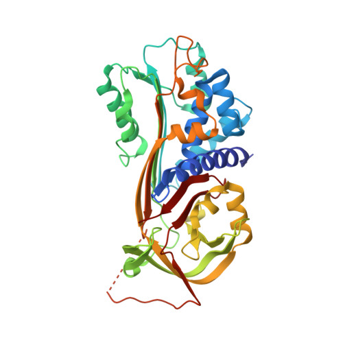 | |
UniProt & NIH Common Fund Data Resources | |||||
Find proteins for P05121 (Homo sapiens) Explore P05121 Go to UniProtKB: P05121 | |||||
PHAROS: P05121 GTEx: ENSG00000106366 | |||||
Entity Groups | |||||
| Sequence Clusters | 30% Identity50% Identity70% Identity90% Identity95% Identity100% Identity | ||||
| UniProt Group | P05121 | ||||
Sequence AnnotationsExpand | |||||
| |||||
| Ligands 1 Unique | |||||
|---|---|---|---|---|---|
| ID | Chains | Name / Formula / InChI Key | 2D Diagram | 3D Interactions | |
| GDE Query on GDE | E [auth A], F [auth C] | 3,4,5-trihydroxybenzoic acid C7 H6 O5 LNTHITQWFMADLM-UHFFFAOYSA-N |  | ||
| Length ( Å ) | Angle ( ˚ ) |
|---|---|
| a = 65.28 | α = 90.91 |
| b = 74.99 | β = 93.29 |
| c = 103.873 | γ = 115.82 |
| Software Name | Purpose |
|---|---|
| HKL-2000 | data collection |
| MOLREP | phasing |
| REFMAC | refinement |
| HKL-2000 | data reduction |
| HKL-2000 | data scaling |
RCSB PDB (citation) is hosted by
RCSB PDB is a member of the

