4HSR

Crystal Structure of a class III engineered cephalosporin acylase


Experimental Data Snapshot

  • Method: X-RAY DIFFRACTION
  • Resolution: 2.13 Å
  • R-Value Free: 0.198 
  • R-Value Work: 0.152 
  • R-Value Observed: 0.154 

wwPDB Validation   3D Report Full Report


This is version 1.3 of the entry. See complete history


Literature

Structure of a class III engineered cephalosporin acylase: comparisons with class I acylase and implications for differences in substrate specificity and catalytic activity.

Golden, E.Paterson, R.Tie, W.J.Anandan, A.Flematti, G.Molla, G.Rosini, E.Pollegioni, L.Vrielink, A.

(2013) Biochem J 451: 217-226

  • DOI: https://doi.org/10.1042/BJ20121715
  • Primary Citation of Related Structures:  
    4HSR, 4HST

  • PubMed Abstract: 

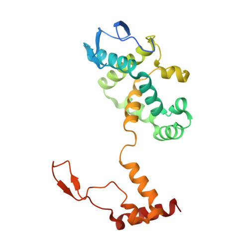
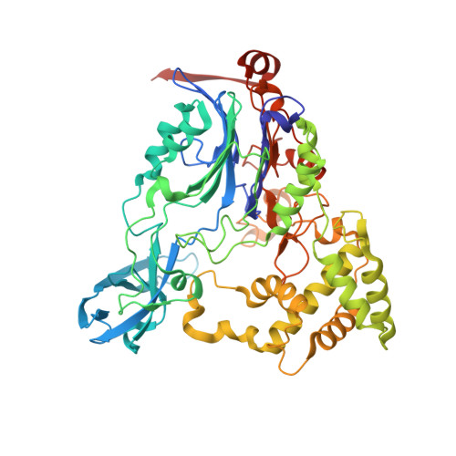
    The crystal structure of the wild-type form of glutaryl-7-ACA (7-aminocephalosporanic acid) acylase from Pseudomonas N176 and a double mutant of the protein (H57βS/H70βS) that displays enhanced catalytic efficiency on cephalosporin C over glutaryl-7-aminocephalosporanic acid has been determined. The structures show a heterodimer made up of an α-chain (229 residues) and a β-chain (543 residues) with a deep cavity, which constitutes the active site. Comparison of the wild-type and mutant structures provides insights into the molecular reasons for the observed enhanced specificity on cephalosporin C over glutaryl-7-aminocephalosporanic acid and offers the basis to evolve a further improved enzyme variant. The nucleophilic catalytic serine residue, Ser(1β), is situated at the base of the active site cavity. The electron density reveals a ligand covalently bound to the catalytic serine residue, such that a tetrahedral adduct is formed. This is proposed to mimic the transition state of the enzyme for both the maturation step and the catalysis of the substrates. A view of the transition state configuration of the enzyme provides important insights into the mechanism of substrate binding and catalysis.


  • Organizational Affiliation

    School of Chemistry and Biochemistry, University of Western Australia, 35 Stirling Highway, Crawley, WA 6009, Australia.


Macromolecules
Find similar proteins by:  (by identity cutoff)  |  3D Structure
Entity ID: 1
MoleculeChains Sequence LengthOrganismDetailsImage
glutaryl-7-aminocephalosporanic acid acylase alpha chain229PseudomonasMutation(s): 0 
Entity Groups  
Sequence Clusters30% Identity50% Identity70% Identity90% Identity95% Identity100% Identity
Sequence Annotations
Expand
  • Reference Sequence
Find similar proteins by:  (by identity cutoff)  |  3D Structure
Entity ID: 2
MoleculeChains Sequence LengthOrganismDetailsImage
glutaryl-7-aminocephalosporanic acid acylase beta chain543PseudomonasMutation(s): 0 
Entity Groups  
Sequence Clusters30% Identity50% Identity70% Identity90% Identity95% Identity100% Identity
Sequence Annotations
Expand
  • Reference Sequence
Small Molecules
Ligands 1 Unique
IDChains Name / Formula / InChI Key2D Diagram3D Interactions
GLJ
Query on GLJ

Download Ideal Coordinates CCD File 
C [auth B]5,5-dihydroxy-L-norvaline
C5 H11 N O4
FIKXYBCGRJPSSD-VKHMYHEASA-N
Experimental Data & Validation

Experimental Data

  • Method: X-RAY DIFFRACTION
  • Resolution: 2.13 Å
  • R-Value Free: 0.198 
  • R-Value Work: 0.152 
  • R-Value Observed: 0.154 
  • Space Group: P 21 21 21
Unit Cell:
Length ( Å )Angle ( ˚ )
a = 68.161α = 90
b = 77.018β = 90
c = 192.138γ = 90
Software Package:
Software NamePurpose
Blu-Icedata collection
SHELXSphasing
REFMACrefinement
XDSdata reduction
SCALAdata scaling

Structure Validation

View Full Validation Report


Entry History 

Deposition Data

Revision History  (Full details and data files)

  • Version 1.0: 2013-02-27
    Type: Initial release
  • Version 1.1: 2013-04-10
    Changes: Database references
  • Version 1.2: 2017-11-15
    Changes: Refinement description
  • Version 1.3: 2024-02-28
    Changes: Data collection, Database references, Derived calculations