4HRM

Structural Basis for Eliciting a Cytotoxic Effect in HER2-Overexpressing Cancer Cells via Binding to the Extracellular Domain of HER2


Experimental Data Snapshot

  • Method: X-RAY DIFFRACTION
  • Resolution: 3.20 Å
  • R-Value Free: 0.339 
  • R-Value Work: 0.314 
  • R-Value Observed: 0.315 

wwPDB Validation   3D Report Full Report


This is version 1.2 of the entry. See complete history


Literature

Structural Basis for Eliciting a Cytotoxic Effect in HER2-Overexpressing Cancer Cells via Binding to the Extracellular Domain of HER2.

Jost, C.Schilling, J.Tamaskovic, R.Schwill, M.Honegger, A.Plueckthun, A.

(2013) Structure 21: 1-13

  • DOI: https://doi.org/10.1016/j.str.2013.08.020
  • Primary Citation of Related Structures:  
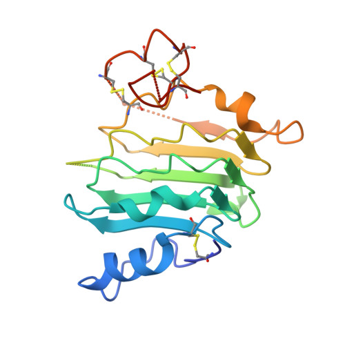
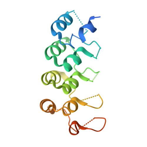
    4HRL, 4HRM, 4HRN

  • PubMed Abstract: 

    Human epidermal growth factor receptor-2 (HER2) is a receptor tyrosine kinase directly linked to the growth of malignancies from various origins and a validated target for monoclonal antibodies and kinase inhibitors. Utilizing a new approach with designed ankyrin repeat proteins (DARPins) as alternative binders, we show that binding of two DARPins connected by a short linker, one targeting extracellular subdomain I and the other subdomain IV, causes much stronger cytotoxic effects on the HER2-addicted breast cancer cell line BT474, surpassing the therapeutic antibody trastuzumab. We determined crystal structures of these DARPins in complex with the respective subdomains. Detailed models of the full-length receptor, constrained by its rigid domain structures and its membrane anchoring, explain how the bispecific DARPins connect two membrane-bound HER2 molecules, distorting them such that they cannot form signaling-competent dimers with any EGFR family member, preventing any kinase dimerization, and thus leading to a complete loss of signaling.


  • Organizational Affiliation

    Department of Biochemistry, University of Zürich, 8057 Zürich, Switzerland.


Macromolecules
Find similar proteins by:  (by identity cutoff)  |  3D Structure
Entity ID: 1
MoleculeChains Sequence LengthOrganismDetailsImage
Domain I of receptor tyrosine-protein kinase erbB-2A [auth C],
B [auth A]
198Homo sapiensMutation(s): 3 
Gene Names: ERBB2HER2MLN19NEUNGL
EC: 2.7.10.1
UniProt & NIH Common Fund Data Resources
Find proteins for P04626 (Homo sapiens)
Explore P04626 
Go to UniProtKB:  P04626
PHAROS:  P04626
GTEx:  ENSG00000141736 
Entity Groups  
Sequence Clusters30% Identity50% Identity70% Identity90% Identity95% Identity100% Identity
UniProt GroupP04626
Sequence Annotations
Expand
  • Reference Sequence
Find similar proteins by:  (by identity cutoff)  |  3D Structure
Entity ID: 2
MoleculeChains Sequence LengthOrganismDetailsImage
Designed Ankyrin Repeat Protein 9_26C [auth B],
D
172synthetic constructMutation(s): 0 
Entity Groups  
Sequence Clusters30% Identity50% Identity70% Identity90% Identity95% Identity100% Identity
Sequence Annotations
Expand
  • Reference Sequence
Experimental Data & Validation

Experimental Data

  • Method: X-RAY DIFFRACTION
  • Resolution: 3.20 Å
  • R-Value Free: 0.339 
  • R-Value Work: 0.314 
  • R-Value Observed: 0.315 
  • Space Group: C 1 2 1
Unit Cell:
Length ( Å )Angle ( ˚ )
a = 138.5α = 90
b = 60.7β = 118.9
c = 107.2γ = 90
Software Package:
Software NamePurpose
XSCALEdata scaling
PHASERphasing
PHENIXrefinement
PDB_EXTRACTdata extraction

Structure Validation

View Full Validation Report


Entry History 

Deposition Data

Revision History  (Full details and data files)

  • Version 1.0: 2013-10-16
    Type: Initial release
  • Version 1.1: 2013-10-30
    Changes: Structure summary
  • Version 1.2: 2023-11-08
    Changes: Data collection, Database references, Refinement description