


Crystal structure of antibiotic biosynthesis monooxygenase
Rice, S., Eswaramoorthy, S., Almo, S.C., Swaminathan, S.To be published.
Experimental Data Snapshot
wwPDB Validation 3D Report Full Report

Entity ID: 1 | |||||
|---|---|---|---|---|---|
| Molecule | Chains | Sequence Length | Organism | Details | Image |
| Antibiotic biosynthesis monooxygenase | 118 | Rhodospirillum rubrum ATCC 11170 | Mutation(s): 0 Gene Names: Rru_A0389 | 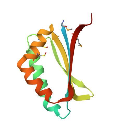 | |
UniProt | |||||
Find proteins for Q2RXF1 (Rhodospirillum rubrum (strain ATCC 11170 / ATH 1.1.1 / DSM 467 / LMG 4362 / NCIMB 8255 / S1)) Explore Q2RXF1 Go to UniProtKB: Q2RXF1 | |||||
Entity Groups | |||||
| Sequence Clusters | 30% Identity50% Identity70% Identity90% Identity95% Identity100% Identity | ||||
| UniProt Group | Q2RXF1 | ||||
Sequence AnnotationsExpand | |||||
| |||||
| Modified Residues 2 Unique | |||||
|---|---|---|---|---|---|
| ID | Chains | Type | Formula | 2D Diagram | Parent |
| CSO Query on CSO | A, B, C, D, E A, B, C, D, E, F, G, H, I | L-PEPTIDE LINKING | C3 H7 N O3 S |  | CYS |
| MSE Query on MSE | A, B, C, D, E A, B, C, D, E, F, G, H, I | L-PEPTIDE LINKING | C5 H11 N O2 Se |  | MET |
| Length ( Å ) | Angle ( ˚ ) |
|---|---|
| a = 176.965 | α = 90 |
| b = 44.535 | β = 101.03 |
| c = 137.913 | γ = 90 |
| Software Name | Purpose |
|---|---|
| CBASS | data collection |
| SHELXS | phasing |
| REFMAC | refinement |
| HKL-2000 | data reduction |
| HKL-2000 | data scaling |
RCSB PDB (citation) is hosted by
RCSB PDB is a member of the

