4H7X

Crystal structure of the tetratricopeptide repeat (TPR) motif of human dual specificity protein kinase Mps1


Experimental Data Snapshot

  • Method: X-RAY DIFFRACTION
  • Resolution: 2.60 Å
  • R-Value Free: 0.240 
  • R-Value Work: 0.193 
  • R-Value Observed: 0.198 

wwPDB Validation   3D Report Full Report


This is version 1.1 of the entry. See complete history


Literature

Structural and functional insights into the role of the N-terminal Mps1 TPR domain in the SAC (spindle assembly checkpoint).

Thebault, P.Chirgadze, D.Y.Dou, Z.Blundell, T.L.Elowe, S.Bolanos-Garcia, V.M.

(2012) Biochem J 448: 321-328

  • DOI: https://doi.org/10.1042/BJ20121448
  • Primary Citation of Related Structures:  
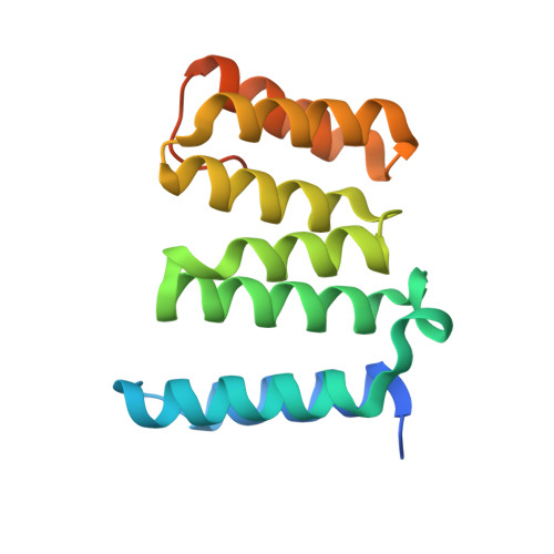
    4H7X, 4H7Y

  • PubMed Abstract: 

    The SAC (spindle assembly checkpoint) is a surveillance system that ensures the timely and accurate transmission of the genetic material to offspring. The process implies kinetochore targeting of the mitotic kinases Bub1 (budding uninhibited by benzamidine 1), BubR1 (Bub1 related) and Mps1 (monopolar spindle 1), which is mediated by the N-terminus of each kinase. In the present study we report the 1.8 Å (1 Å=0.1 nm) crystal structure of the TPR (tetratricopeptide repeat) domain in the N-terminal region of human Mps1. The structure reveals an overall high similarity to the TPR motif of the mitotic checkpoint kinases Bub1 and BubR1, and a number of unique features that include the absence of the binding site for the kinetochore structural component KNL1 (kinetochore-null 1; blinkin), and determinants of dimerization. Moreover, we show that a stretch of amino acids at the very N-terminus of Mps1 is required for dimer formation, and that interfering with dimerization results in mislocalization and misregulation of kinase activity. The results of the present study provide an important insight into the molecular details of the mitotic functions of Mps1 including features that dictate substrate selectivity and kinetochore docking.


  • Organizational Affiliation

    Centre de Recherche du CHUQ, 2705 Boulevard Laurier, RC-9800 Québec, Canada G1V 4G2.


Macromolecules
Find similar proteins by:  (by identity cutoff)  |  3D Structure
Entity ID: 1
MoleculeChains Sequence LengthOrganismDetailsImage
Dual specificity protein kinase TTK
A, B
161Homo sapiensMutation(s): 0 
Gene Names: MPS1MPS1L1NCBI Reference Sequence NM_003318.4TTK
EC: 2.7.12.1
UniProt & NIH Common Fund Data Resources
Find proteins for P33981 (Homo sapiens)
Explore P33981 
Go to UniProtKB:  P33981
PHAROS:  P33981
GTEx:  ENSG00000112742 
Entity Groups  
Sequence Clusters30% Identity50% Identity70% Identity90% Identity95% Identity100% Identity
UniProt GroupP33981
Sequence Annotations
Expand
  • Reference Sequence
Small Molecules
Ligands 1 Unique
IDChains Name / Formula / InChI Key2D Diagram3D Interactions
PB
Query on PB

Download Ideal Coordinates CCD File 
C [auth A]LEAD (II) ION
Pb
RVPVRDXYQKGNMQ-UHFFFAOYSA-N
Experimental Data & Validation

Experimental Data

  • Method: X-RAY DIFFRACTION
  • Resolution: 2.60 Å
  • R-Value Free: 0.240 
  • R-Value Work: 0.193 
  • R-Value Observed: 0.198 
  • Space Group: P 41 21 2
Unit Cell:
Length ( Å )Angle ( ˚ )
a = 79.487α = 90
b = 79.487β = 90
c = 137.684γ = 90
Software Package:
Software NamePurpose
GDAdata collection
PHENIXmodel building
PHENIXrefinement
DENZOdata reduction
SCALEPACKdata scaling
PHENIXphasing

Structure Validation

View Full Validation Report


Entry History 

Deposition Data

Revision History  (Full details and data files)

  • Version 1.0: 2012-12-12
    Type: Initial release
  • Version 1.1: 2024-02-28
    Changes: Data collection, Database references, Derived calculations