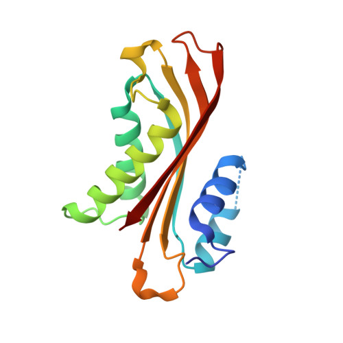
4H4L

Crystal Structure of ternary complex of HutP(HutP-L-His-Zn)


Experimental Data Snapshot

  • Method: X-RAY DIFFRACTION
  • Resolution: 2.50 Å
  • R-Value Free: 0.298 
  • R-Value Work: 0.219 
  • R-Value Observed: 0.223 

wwPDB Validation   3D Report Full Report


Ligand Structure Quality Assessment 


This is version 1.3 of the entry. See complete history


Literature

Alternative binding modes of l-histidine guided by metal ions for the activation of the antiterminator protein HutP of Bacillus subtilis.

Dhakshnamoorthy, B.Mizuno, H.Kumar, P.K.R.

(2013) J Struct Biol 183: 512-518

  • DOI: https://doi.org/10.1016/j.jsb.2013.05.019
  • Primary Citation of Related Structures:  
    4H4L

  • PubMed Abstract: 

    Anti-terminator proteins control gene expression by recognizing control signals within cognate transcripts and then preventing transcription termination. HutP is such a regulatory protein that regulates the expression of the histidine utilization (hut) operon in Bacillus subtilis by binding to cis-acting regulatory sequences in hut mRNAs. During the anti-termination process, l-histidine and a divalent ion are required for hutP to bind to the specific sequence within the hut mRNA. Our previous crystal structure of the HutP-l-histidine-Mg(2+)-RNA ternary complex demonstrated that the l-histidine ligand and Mg(2+) bind together such that the backbone nitrogen and carboxyl oxygen of l-histidine coordinate with Mg(2+). In addition to the Mg(2+), other divalent ions are also known to efficiently support the l-histidine-dependent anti-termination of the hut operon, and the best divalent ion is Zn(2+). In this study, we determined the crystal structure of the HutP-l-histidine-Zn(2+) complex and found that the orientation of l-histidine coordinated to Zn(2+) is reversed relative to that of l-histidine coordinated to Mg(2+), i.e., the imidazole side chain nitrogen of l-histidine coordinates to Zn(2+). This alternative binding mode of the l-histidine ligand to a divalent ion provides further insight into the mechanisms responsible for the activation of RNA binding during the hut anti-termination process.


  • Organizational Affiliation

    RNA Processing Group, Biomedical Research Institute, National Institute of Advanced Industrial Science and Technology, Central-6, 1-1-1 Higashi, Tsukuba 305-8566, Japan.


Macromolecules
Find similar proteins by:  (by identity cutoff)  |  3D Structure
Entity ID: 1
MoleculeChains Sequence LengthOrganismDetailsImage
Hut operon positive regulatory protein
A, B, C, D, E
A, B, C, D, E, F, G, H, I, J, K, L
148Bacillus subtilis subsp. subtilis str. 168Mutation(s): 1 
Gene Names: BSU39340hutP
UniProt
Find proteins for P10943 (Bacillus subtilis (strain 168))
Explore P10943 
Go to UniProtKB:  P10943
Entity Groups  
Sequence Clusters30% Identity50% Identity70% Identity90% Identity95% Identity100% Identity
UniProt GroupP10943
Sequence Annotations
Expand
  • Reference Sequence
Small Molecules
Ligands 2 Unique
IDChains Name / Formula / InChI Key2D Diagram3D Interactions
HIS
Query on HIS

Download Ideal Coordinates CCD File 
AA [auth G]
CA [auth I]
EA [auth J]
GA [auth J]
IA [auth L]
AA [auth G],
CA [auth I],
EA [auth J],
GA [auth J],
IA [auth L],
M [auth A],
O [auth B],
Q [auth C],
S [auth D],
U [auth D],
W [auth F],
Y [auth G]
HISTIDINE
C6 H10 N3 O2
HNDVDQJCIGZPNO-YFKPBYRVSA-O
ZN
Query on ZN

Download Ideal Coordinates CCD File 
BA [auth H]
DA [auth I]
FA [auth J]
HA [auth K]
JA [auth L]
BA [auth H],
DA [auth I],
FA [auth J],
HA [auth K],
JA [auth L],
N [auth A],
P [auth B],
R [auth C],
T [auth D],
V [auth E],
X [auth F],
Z [auth G]
ZINC ION
Zn
PTFCDOFLOPIGGS-UHFFFAOYSA-N
Experimental Data & Validation

Experimental Data

  • Method: X-RAY DIFFRACTION
  • Resolution: 2.50 Å
  • R-Value Free: 0.298 
  • R-Value Work: 0.219 
  • R-Value Observed: 0.223 
  • Space Group: P 3
Unit Cell:
Length ( Å )Angle ( ˚ )
a = 129.53α = 90
b = 129.53β = 90
c = 76.67γ = 120
Software Package:
Software NamePurpose
ADSCdata collection
PHASERphasing
REFMACrefinement
HKL-2000data reduction
HKL-2000data scaling

Structure Validation

View Full Validation Report


Ligand Structure Quality Assessment 


Entry History 

Deposition Data

Revision History  (Full details and data files)

  • Version 1.0: 2013-09-04
    Type: Initial release
  • Version 1.1: 2014-09-10
    Changes: Database references
  • Version 1.2: 2017-11-15
    Changes: Advisory, Refinement description
  • Version 1.3: 2023-11-08
    Changes: Advisory, Data collection, Database references, Derived calculations, Refinement description