4H36

Crystal Structure of JNK3 in Complex with ATF2 Peptide


Experimental Data Snapshot

  • Method: X-RAY DIFFRACTION
  • Resolution: 3.00 Å
  • R-Value Free: 0.269 
  • R-Value Work: 0.216 
  • R-Value Observed: 0.219 

wwPDB Validation   3D Report Full Report


This is version 1.5 of the entry. See complete history


Literature

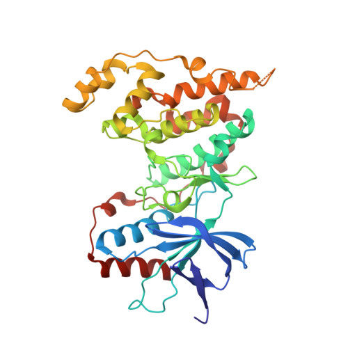
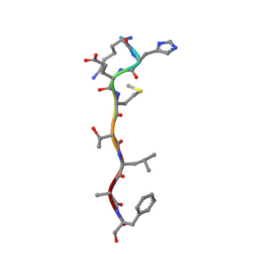
Structural Mechanisms of Allostery and Autoinhibition in JNK Family Kinases.

Laughlin, J.D.Nwachukwu, J.C.Figuera-Losada, M.Cherry, L.Nettles, K.W.Lograsso, P.V.

(2012) Structure 20: 2174-2184

  • DOI: https://doi.org/10.1016/j.str.2012.09.021
  • Primary Citation of Related Structures:  
    4H36, 4H39, 4H3B

  • PubMed Abstract: 

    c-Jun N-terminal (JNK) family kinases have a common peptide-docking site used by upstream activating kinases, substrates, scaffold proteins, and phosphatases, where the ensemble of bound proteins determines signaling output. Although there are many JNK structures, little is known about mechanisms of allosteric regulation between the catalytic and peptide-binding sites, and the activation loop, whose phosphorylation is required for catalytic activity. Here, we compare three structures of unliganded JNK3 bound to different peptides. These were compared as a class to structures that differ in binding of peptide, small molecule ligand, or conformation of the kinase activation loop. Peptide binding induced an inhibitory interlobe conformer that was reversed by alterations in the activation loop. Structure class analysis revealed the subtle structural mechanisms for allosteric signaling between the peptide-binding site and activation loop. Biochemical data from isothermal calorimetry, fluorescence energy transfer, and enzyme inhibition demonstrated affinity differences among the three peptides that were consistent with structural observations.


  • Organizational Affiliation

    Department of Molecular Therapeutics, The Scripps Research Institute, 130 Scripps Way, Jupiter, FL 33458, USA.


Macromolecules
Find similar proteins by:  (by identity cutoff)  |  3D Structure
Entity ID: 1
MoleculeChains Sequence LengthOrganismDetailsImage
Mitogen-activated protein kinase 10356Homo sapiensMutation(s): 0 
Gene Names: JNK3JNK3AMAPK10PRKM10SAPK1B
EC: 2.7.11.24
UniProt & NIH Common Fund Data Resources
Find proteins for P53779 (Homo sapiens)
Explore P53779 
Go to UniProtKB:  P53779
PHAROS:  P53779
GTEx:  ENSG00000109339 
Entity Groups  
Sequence Clusters30% Identity50% Identity70% Identity90% Identity95% Identity100% Identity
UniProt GroupP53779
Sequence Annotations
Expand
  • Reference Sequence

Find similar proteins by:  Sequence   |   3D Structure  

Entity ID: 2
MoleculeChains Sequence LengthOrganismDetailsImage
Cyclic AMP-dependent transcription factor ATF-28Homo sapiensMutation(s): 0 
Gene Names: ATF2CREB2CREBP1
UniProt & NIH Common Fund Data Resources
Find proteins for P15336 (Homo sapiens)
Explore P15336 
Go to UniProtKB:  P15336
PHAROS:  P15336
GTEx:  ENSG00000115966 
Entity Groups  
UniProt GroupP15336
Sequence Annotations
Expand
  • Reference Sequence
Experimental Data & Validation

Experimental Data

  • Method: X-RAY DIFFRACTION
  • Resolution: 3.00 Å
  • R-Value Free: 0.269 
  • R-Value Work: 0.216 
  • R-Value Observed: 0.219 
  • Space Group: P 31 2 1
Unit Cell:
Length ( Å )Angle ( ˚ )
a = 84.58α = 90
b = 84.58β = 90
c = 127.06γ = 120
Software Package:
Software NamePurpose
Blu-Icedata collection
PHENIXmodel building
PHENIXrefinement
HKL-2000data reduction
HKL-2000data scaling
PHENIXphasing

Structure Validation

View Full Validation Report


Entry History 

Deposition Data

Revision History  (Full details and data files)

  • Version 1.0: 2012-11-21
    Type: Initial release
  • Version 1.1: 2012-11-28
    Changes: Database references
  • Version 1.2: 2012-12-26
    Changes: Database references
  • Version 1.3: 2015-06-17
    Changes: Structure summary
  • Version 1.4: 2017-11-15
    Changes: Refinement description
  • Version 1.5: 2023-09-20
    Changes: Data collection, Database references, Refinement description