4GVB

Crystal structure of the virally encoded antifungal protein, KP6, heterodimer


Experimental Data Snapshot

  • Method: X-RAY DIFFRACTION
  • Resolution: 1.80 Å
  • R-Value Free: 0.247 
  • R-Value Work: 0.215 
  • R-Value Observed: 0.216 

wwPDB Validation   3D Report Full Report


This is version 1.2 of the entry. See complete history


Literature

The Atomic Structure of the Virally Encoded Antifungal Protein, KP6.

Allen, A.Chatt, E.Smith, T.J.

(2013) J Mol Biol 425: 609-621

  • DOI: https://doi.org/10.1016/j.jmb.2012.11.033
  • Primary Citation of Related Structures:  
    4GVB

  • PubMed Abstract: 

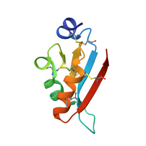
    Killer toxins are produced by several genera of yeast and filamentous fungi. A small proportion of Ustilago maydis strains produce killer toxins, to which they are resistant, but sensitive strains are the majority in the wild populations. There are three killer types (P1, P4 and P6) that secrete KP1, KP4 and KP6 toxins, respectively, which are produced only by strains persistently infected with double-stranded RNA viruses (UmV) in the cell cytoplasm. Unlike nearly all other viruses, UmV are only transmitted through mitosis or meiosis. As shown here, KP6 is different from any other known cytotoxic protein. KP6 is neutral protein composed of two subunits: KP6α and KP6β. KP6α is responsible for targeting while KP6β is cytotoxic. Neither subunit is homologous in either sequence or structure to any other toxin, but they have highly similar structures to each other. The major difference between the two subunits is a hydrophobic helix at the N-terminus of KP6α and is likely key to target recognition. Unlike any other toxin, KP6 is translated as a single polypeptide with a 31-residue linker region in the middle of the protein. From structural prediction studies, this linker likely makes for a more compact KP6 structure that sequesters the hydrophobic helix of KP6α. A model whereby the protoxin undergoes a conformational activation process that exposes this helix immediately prior to secretion is presented.


  • Organizational Affiliation

    Donald Danforth Plant Science Center, 975 North Warson Road, St. Louis, MO 63132, USA.


Macromolecules
Find similar proteins by:  (by identity cutoff)  |  3D Structure
Entity ID: 1
MoleculeChains Sequence LengthOrganismDetailsImage
KP6 killer toxin subunit alpha80Ustilago maydis virus P6Mutation(s): 0 
UniProt
Find proteins for P16948 (Ustilago maydis P6 virus)
Explore P16948 
Go to UniProtKB:  P16948
Entity Groups  
Sequence Clusters30% Identity50% Identity70% Identity90% Identity95% Identity100% Identity
UniProt GroupP16948
Sequence Annotations
Expand
  • Reference Sequence
Find similar proteins by:  (by identity cutoff)  |  3D Structure
Entity ID: 2
MoleculeChains Sequence LengthOrganismDetailsImage
KP6 killer toxin subunit beta81Ustilago maydis virus P6Mutation(s): 0 
UniProt
Find proteins for P16948 (Ustilago maydis P6 virus)
Explore P16948 
Go to UniProtKB:  P16948
Entity Groups  
Sequence Clusters30% Identity50% Identity70% Identity90% Identity95% Identity100% Identity
UniProt GroupP16948
Sequence Annotations
Expand
  • Reference Sequence
Experimental Data & Validation

Experimental Data

  • Method: X-RAY DIFFRACTION
  • Resolution: 1.80 Å
  • R-Value Free: 0.247 
  • R-Value Work: 0.215 
  • R-Value Observed: 0.216 
  • Space Group: P 32 2 1
Unit Cell:
Length ( Å )Angle ( ˚ )
a = 44.419α = 90
b = 44.419β = 90
c = 155.648γ = 120
Software Package:
Software NamePurpose
PROTEUM PLUSdata collection
MOLREPphasing
PHENIXrefinement
SAINTdata reduction
PROTEUM PLUSdata scaling

Structure Validation

View Full Validation Report


Entry History 

Deposition Data

  • Released Date: 2012-12-19 
  • Deposition Author(s): Smith, T.

Revision History  (Full details and data files)

  • Version 1.0: 2012-12-19
    Type: Initial release
  • Version 1.1: 2013-01-23
    Changes: Database references
  • Version 1.2: 2013-02-13
    Changes: Database references