


4GKO
Crystal structure of the calcium2+-bound human IgE-Fc(epsilon)3-4 bound to its B cell receptor derCD23
- PDB DOI: https://doi.org/10.2210/pdb4GKO/pdb
- Classification: IMMUNE SYSTEM
- Organism(s): Homo sapiens
- Expression System: Homo sapiens, Escherichia coli
- Mutation(s): No
- Deposited: 2012-08-13 Released: 2013-06-26
Experimental Data Snapshot
- Method: X-RAY DIFFRACTION
- Resolution: 3.30 Å
- R-Value Free: 0.312
- R-Value Work: 0.273
- R-Value Observed: 0.275

This is version 2.1 of the entry. See complete history.

Macromolecules
Find similar proteins by:
(by identity cutoff) | 3D Structure
Entity ID: 1 | |||||
|---|---|---|---|---|---|
| Molecule | Chains | Sequence Length | Organism | Details | Image |
| Ig epsilon chain C region | 223 | Homo sapiens | Mutation(s): 0 Gene Names: IGHE | 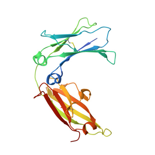 | |
UniProt & NIH Common Fund Data Resources | |||||
Find proteins for P01854 (Homo sapiens) Explore P01854 Go to UniProtKB: P01854 | |||||
PHAROS: P01854 GTEx: ENSG00000211891 | |||||
Entity Groups | |||||
| Sequence Clusters | 30% Identity50% Identity70% Identity90% Identity95% Identity100% Identity | ||||
| UniProt Group | P01854 | ||||
Sequence AnnotationsExpand | |||||
| |||||
Find similar proteins by:
(by identity cutoff) | 3D Structure
Entity ID: 2 | |||||
|---|---|---|---|---|---|
| Molecule | Chains | Sequence Length | Organism | Details | Image |
| Low affinity immunoglobulin epsilon Fc receptor | 143 | Homo sapiens | Mutation(s): 0 Gene Names: FCER2, CD23A, CLEC4J, FCE2, IGEBF | 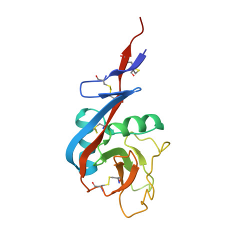 | |
UniProt & NIH Common Fund Data Resources | |||||
Find proteins for P06734 (Homo sapiens) Explore P06734 Go to UniProtKB: P06734 | |||||
PHAROS: P06734 GTEx: ENSG00000104921 | |||||
Entity Groups | |||||
| Sequence Clusters | 30% Identity50% Identity70% Identity90% Identity95% Identity100% Identity | ||||
| UniProt Group | P06734 | ||||
Sequence AnnotationsExpand | |||||
| |||||
Oligosaccharides
Entity ID: 3 | |||||
|---|---|---|---|---|---|
| Molecule | Chains | Length | 2D Diagram | Glycosylation | 3D Interactions |
| alpha-D-mannopyranose-(1-3)-[alpha-D-mannopyranose-(1-6)]beta-D-mannopyranose-(1-4)-2-acetamido-2-deoxy-beta-D-glucopyranose-(1-4)-2-acetamido-2-deoxy-beta-D-glucopyranose | M, N, O, P, Q M, N, O, P, Q, R | 5 |  | N/A | |
Glycosylation Resources | |||||
GlyTouCan: G22768VO GlyCosmos: G22768VO GlyGen: G22768VO | |||||
Small Molecules
| Ligands 2 Unique | |||||
|---|---|---|---|---|---|
| ID | Chains | Name / Formula / InChI Key | 2D Diagram | 3D Interactions | |
| MAN Query on MAN | S [auth C], T [auth C] | alpha-D-mannopyranose C6 H12 O6 WQZGKKKJIJFFOK-PQMKYFCFSA-N |  | ||
| CA Query on CA | U [auth G] V [auth H] W [auth I] X [auth J] Y [auth K] | CALCIUM ION Ca BHPQYMZQTOCNFJ-UHFFFAOYSA-N |  | ||
Experimental Data & Validation
Experimental Data
- Method: X-RAY DIFFRACTION
- Resolution: 3.30 Å
- R-Value Free: 0.312
- R-Value Work: 0.273
- R-Value Observed: 0.275
- Space Group: P 21 21 21
Unit Cell:
| Length ( Å ) | Angle ( ˚ ) |
|---|---|
| a = 62.83 | α = 90 |
| b = 110.13 | β = 90 |
| c = 367.41 | γ = 90 |
| Software Name | Purpose |
|---|---|
| REFMAC | refinement |
| PDB_EXTRACT | data extraction |
| HKL-2000 | data collection |
| MOSFLM | data reduction |
| SCALA | data scaling |
| PHASES | phasing |
Entry History
Deposition Data
- Released Date: 2013-06-26 Deposition Author(s): Yuan, D., Sutton, B.J., Dhaliwal, B.
Revision History (Full details and data files)
- Version 1.0: 2013-06-26
Type: Initial release - Version 1.1: 2013-08-14
Changes: Database references - Version 2.0: 2020-07-29
Type: Remediation
Reason: Carbohydrate remediation
Changes: Advisory, Atomic model, Data collection, Database references, Derived calculations, Structure summary - Version 2.1: 2023-09-13
Changes: Data collection, Database references, Refinement description, Structure summary
