4GF9

Structural insights into the dual strategy of recognition of peptidoglycan recognition protein, PGRP-S: ternary complex of PGRP-S with LPS and fatty acid


Experimental Data Snapshot

  • Method: X-RAY DIFFRACTION
  • Resolution: 2.80 Å
  • R-Value Free: 0.265 
  • R-Value Work: 0.227 
  • R-Value Observed: 0.229 

wwPDB Validation   3D Report Full Report


Ligand Structure Quality Assessment 


This is version 1.2 of the entry. See complete history


Literature

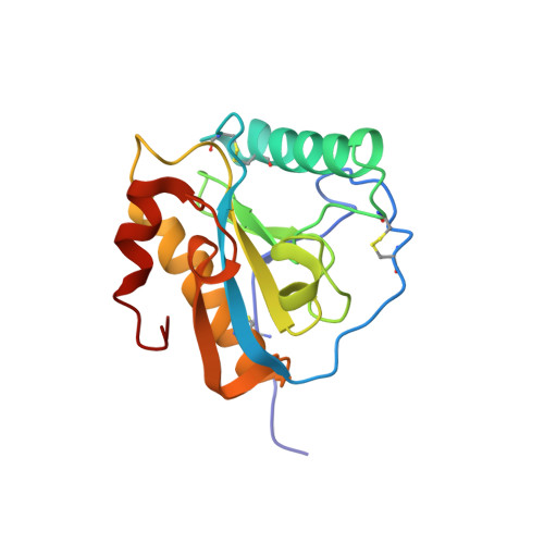
Structural insights into the dual strategy of recognition by peptidoglycan recognition protein, PGRP-S: structure of the ternary complex of PGRP-S with lipopolysaccharide and stearic acid.

Sharma, P.Dube, D.Sinha, M.Yadav, S.Kaur, P.Sharma, S.Singh, T.P.

(2013) PLoS One 8: e53756-e53756

  • DOI: https://doi.org/10.1371/journal.pone.0053756
  • Primary Citation of Related Structures:  
    4GF9

  • PubMed Abstract: 

    Peptidoglycan recognition proteins (PGRPs) are part of the innate immune system. The 19 kDa Short PGRP (PGRP-S) is one of the four mammalian PGRPs. The concentration of PGRP-S in camel (CPGRP-S) has been shown to increase considerably during mastitis. The structure of CPGRP-S consists of four protein molecules designated as A, B, C and D forming stable intermolecular contacts, A-B and C-D. The A-B and C-D interfaces are located on the opposite sides of the same monomer leading to the the formation of a linear chain with alternating A-B and C-D contacts. Two ligand binding sites, one at C-D contact and another at A-B contact have been observed. CPGRP-S binds to the components of bacterial cell wall molecules such as lipopolysaccharide (LPS), lipoteichoic acid (LTA), and peptidoglycan (PGN) from both gram-positive and gram-negative bacteria. It also binds to fatty acids including mycolic acid of the Mycobacterium tuberculosis (Mtb). Previous structural studies of binary complexes of CPGRP-S with LPS and stearic acid (SA) have shown that LPS binds to CPGRP-S at C-D contact (Site-1) while SA binds to it at the A-B contact (Site-2). The binding studies using surface plasmon resonance showed that LPS and SA bound to CPGRP-S in the presence of each other. The structure determination of the ternary complex showed that LPS and SA bound to CPGRP-S at Site-1 and Site-2 respectively. LPS formed 13 hydrogen bonds and 159 van der Waals contacts (distances ≤4.2 Å) while SA formed 56 van der Waals contacts. The ELISA test showed that increased levels of productions of pro-inflammatory cytokines TNF-α and IFN-γ due to LPS and SA decreased considerably upon the addition of CPGRP-S.


  • Organizational Affiliation

    Department of Biophysics, All India Institute of Medical Sciences, New Delhi, India.


Macromolecules
Find similar proteins by:  (by identity cutoff)  |  3D Structure
Entity ID: 1
MoleculeChains Sequence LengthOrganismDetailsImage
Peptidoglycan recognition protein 1
A, B, C, D
171Camelus dromedariusMutation(s): 0 
UniProt
Find proteins for Q9GK12 (Camelus dromedarius)
Explore Q9GK12 
Go to UniProtKB:  Q9GK12
Entity Groups  
Sequence Clusters30% Identity50% Identity70% Identity90% Identity95% Identity100% Identity
UniProt GroupQ9GK12
Sequence Annotations
Expand
  • Reference Sequence
Small Molecules
Ligands 3 Unique
IDChains Name / Formula / InChI Key2D Diagram3D Interactions
LP5
Query on LP5

Download Ideal Coordinates CCD File 
F [auth C](R)-((2R,3S,4R,5R,6R)-3-HYDROXY-2-(HYDROXYMETHYL)-5-((R)-3-HYDROXYTETRADECANAMIDO)-6-(PHOSPHONOOXY)TETRAHYDRO-2H-PYRAN-4-YL) 3-HYDROXYTETRADECANOATE
C34 H66 N O12 P
HEHQDWUWJVPREQ-XQJZMFRCSA-N
STE
Query on STE

Download Ideal Coordinates CCD File 
E [auth B]STEARIC ACID
C18 H36 O2
QIQXTHQIDYTFRH-UHFFFAOYSA-N
GOL
Query on GOL

Download Ideal Coordinates CCD File 
G [auth D]GLYCEROL
C3 H8 O3
PEDCQBHIVMGVHV-UHFFFAOYSA-N
Experimental Data & Validation

Experimental Data

  • Method: X-RAY DIFFRACTION
  • Resolution: 2.80 Å
  • R-Value Free: 0.265 
  • R-Value Work: 0.227 
  • R-Value Observed: 0.229 
  • Space Group: I 2 2 2
Unit Cell:
Length ( Å )Angle ( ˚ )
a = 89.62α = 90
b = 101.88β = 90
c = 163.313γ = 90
Software Package:
Software NamePurpose
HKL-2000data collection
AMoREphasing
REFMACrefinement
HKL-2000data reduction
HKL-2000data scaling

Structure Validation

View Full Validation Report


Ligand Structure Quality Assessment 


Entry History 

Deposition Data

Revision History  (Full details and data files)

  • Version 1.0: 2012-09-26
    Type: Initial release
  • Version 1.1: 2013-05-15
    Changes: Database references
  • Version 1.2: 2023-09-13
    Changes: Data collection, Database references, Derived calculations, Refinement description