4GBX

Crystal structure of an immune complex at pH 6.5


Experimental Data Snapshot

  • Method: X-RAY DIFFRACTION
  • Resolution: 3.00 Å
  • R-Value Free: 0.250 
  • R-Value Work: 0.198 
  • R-Value Observed: 0.200 

wwPDB Validation   3D Report Full Report


Ligand Structure Quality Assessment 


This is version 1.2 of the entry. See complete history


Literature

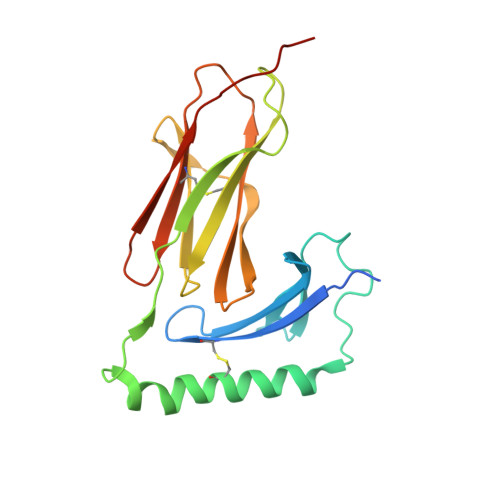
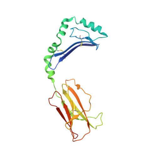
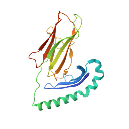
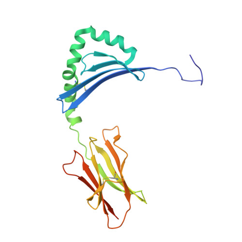
Crystal Structure of the HLA-DM-HLA-DR1 Complex Defines Mechanisms for Rapid Peptide Selection.

Pos, W.Sethi, D.K.Call, M.J.Schulze, M.S.Anders, A.K.Pyrdol, J.Wucherpfennig, K.W.

(2012) Cell 151: 1557-1568

  • DOI: https://doi.org/10.1016/j.cell.2012.11.025
  • Primary Citation of Related Structures:  
    4FQX, 4GBX

  • PubMed Abstract: 

    HLA-DR molecules bind microbial peptides in an endosomal compartment and present them on the cell surface for CD4 T cell surveillance. HLA-DM plays a critical role in the endosomal peptide selection process. The structure of the HLA-DM-HLA-DR complex shows major rearrangements of the HLA-DR peptide-binding groove. Flipping of a tryptophan away from the HLA-DR1 P1 pocket enables major conformational changes that position hydrophobic HLA-DR residues into the P1 pocket. These conformational changes accelerate peptide dissociation and stabilize the empty HLA-DR peptide-binding groove. Initially, incoming peptides have access to only part of the HLA-DR groove and need to compete with HLA-DR residues for access to the P2 site and the hydrophobic P1 pocket. This energetic barrier creates a rapid and stringent selection process for the highest-affinity binders. Insertion of peptide residues into the P2 and P1 sites reverses the conformational changes, terminating selection through DM dissociation.


  • Organizational Affiliation

    Department of Cancer Immunology & AIDS, Dana-Farber Cancer Institute, Boston, MA 02115, USA.


Macromolecules
Find similar proteins by:  (by identity cutoff)  |  3D Structure
Entity ID: 1
MoleculeChains Sequence LengthOrganismDetailsImage
HLA class II histocompatibility antigen, DM alpha chainA [auth C]203Homo sapiensMutation(s): 3 
Gene Names: alpha chainDMAHLA-DMHLA-DMAMHC CLASS II MOLECULERING6
UniProt & NIH Common Fund Data Resources
Find proteins for P28067 (Homo sapiens)
Explore P28067 
Go to UniProtKB:  P28067
PHAROS:  P28067
GTEx:  ENSG00000204257 
Entity Groups  
Sequence Clusters30% Identity50% Identity70% Identity90% Identity95% Identity100% Identity
UniProt GroupP28067
Sequence Annotations
Expand
  • Reference Sequence
Find similar proteins by:  (by identity cutoff)  |  3D Structure
Entity ID: 2
MoleculeChains Sequence LengthOrganismDetailsImage
HLA class II histocompatibility antigen, DM beta chainB [auth D]199Homo sapiensMutation(s): 2 
Gene Names: beta chainDMBHLA-DMBMHC Class II molecule HLA-DMRING7
UniProt & NIH Common Fund Data Resources
Find proteins for P28068 (Homo sapiens)
Explore P28068 
Go to UniProtKB:  P28068
PHAROS:  P28068
GTEx:  ENSG00000242574 
Entity Groups  
Sequence Clusters30% Identity50% Identity70% Identity90% Identity95% Identity100% Identity
UniProt GroupP28068
Sequence Annotations
Expand
  • Reference Sequence
Find similar proteins by:  (by identity cutoff)  |  3D Structure
Entity ID: 3
MoleculeChains Sequence LengthOrganismDetailsImage
HLA class II histocompatibility antigen, DR alpha chainC [auth A]191Homo sapiensMutation(s): 1 
Gene Names: alpha chainHLA-DR1HLA-DRAHLA-DRA1MHC Class II molecule
UniProt & NIH Common Fund Data Resources
Find proteins for P01903 (Homo sapiens)
Explore P01903 
Go to UniProtKB:  P01903
PHAROS:  P01903
GTEx:  ENSG00000204287 
Entity Groups  
Sequence Clusters30% Identity50% Identity70% Identity90% Identity95% Identity100% Identity
UniProt GroupP01903
Sequence Annotations
Expand
  • Reference Sequence
Find similar proteins by:  (by identity cutoff)  |  3D Structure
Entity ID: 4
MoleculeChains Sequence LengthOrganismDetailsImage
HLA class II histocompatibility antigen, DRB1-1 beta chainD [auth B]208Homo sapiensMutation(s): 1 
Gene Names: beta chainHLA-DR1HLA-DRB1MHC Class II molecule
UniProt & NIH Common Fund Data Resources
Find proteins for P01911 (Homo sapiens)
Explore P01911 
Go to UniProtKB:  P01911
PHAROS:  P01911
GTEx:  ENSG00000196126 
Entity Groups  
Sequence Clusters30% Identity50% Identity70% Identity90% Identity95% Identity100% Identity
UniProt GroupP01911
Sequence Annotations
Expand
  • Reference Sequence

Find similar proteins by:  Sequence   |   3D Structure  

Entity ID: 5
MoleculeChains Sequence LengthOrganismDetailsImage
synthetic peptide10synthetic constructMutation(s): 0 
Entity Groups  
Sequence Clusters30% Identity50% Identity70% Identity90% Identity95% Identity100% Identity
Sequence Annotations
Expand
  • Reference Sequence
Experimental Data & Validation

Experimental Data

  • Method: X-RAY DIFFRACTION
  • Resolution: 3.00 Å
  • R-Value Free: 0.250 
  • R-Value Work: 0.198 
  • R-Value Observed: 0.200 
  • Space Group: P 21 21 21
Unit Cell:
Length ( Å )Angle ( ˚ )
a = 66.167α = 90
b = 126.032β = 90
c = 143.107γ = 90
Software Package:
Software NamePurpose
SCALEPACKdata scaling
PHASERphasing
PHENIXrefinement
PDB_EXTRACTdata extraction
ADSCdata collection
DENZOdata reduction

Structure Validation

View Full Validation Report


Ligand Structure Quality Assessment 


Entry History 

Deposition Data

Revision History  (Full details and data files)

  • Version 1.0: 2013-01-16
    Type: Initial release
  • Version 1.1: 2017-11-15
    Changes: Refinement description
  • Version 1.2: 2020-07-29
    Type: Remediation
    Reason: Carbohydrate remediation
    Changes: Data collection, Database references, Derived calculations, Structure summary