


Crystal structure of Eph receptor 5
Shi, J.H., Zhu, W.L., Song, J.X.To be published.
Experimental Data Snapshot
wwPDB Validation 3D Report Full Report

Entity ID: 1 | |||||
|---|---|---|---|---|---|
| Molecule | Chains | Sequence Length | Organism | Details | Image |
| Ephrin type-A receptor 5 | 179 | Homo sapiens | Mutation(s): 1 Gene Names: BSK, EHK1, EPHA5, HEK7, TYRO4 EC: 2.7.10.1 | 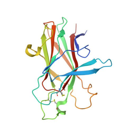 | |
UniProt & NIH Common Fund Data Resources | |||||
Find proteins for P54756 (Homo sapiens) Explore P54756 Go to UniProtKB: P54756 | |||||
PHAROS: P54756 GTEx: ENSG00000145242 | |||||
Entity Groups | |||||
| Sequence Clusters | 30% Identity50% Identity70% Identity90% Identity95% Identity100% Identity | ||||
| UniProt Group | P54756 | ||||
Sequence AnnotationsExpand | |||||
| |||||
| Length ( Å ) | Angle ( ˚ ) |
|---|---|
| a = 55.04 | α = 90 |
| b = 82.721 | β = 90 |
| c = 81.166 | γ = 90 |
| Software Name | Purpose |
|---|---|
| PHENIX | refinement |
| PROTEUM PLUS | data collection |
| SAINT | data reduction |
| PROTEUM PLUS | data scaling |
| PHASES | phasing |
RCSB PDB (citation) is hosted by
RCSB PDB is a member of the

