4EF8

Crystal structure of dihydroorotate dehydrogenase from Leishmania major in complex with Phenyl isothiocyanate


Experimental Data Snapshot

  • Method: X-RAY DIFFRACTION
  • Resolution: 1.56 Å
  • R-Value Free: 0.194 
  • R-Value Work: 0.176 
  • R-Value Observed: 0.177 

wwPDB Validation   3D Report Full Report


Ligand Structure Quality Assessment 


This is version 1.2 of the entry. See complete history


Literature

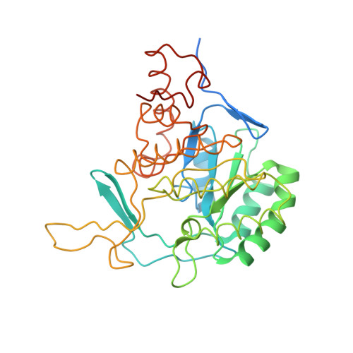
Target sites for the design of anti-trypanosomatid drugs based on the structure of dihydroorotate dehydrogenase.

Pinheiro, M.P.Emery, F.S.Nonato, M.C.

(2013) Curr Pharm Des 19: 2615-2627

  • DOI: https://doi.org/10.2174/1381612811319140011
  • Primary Citation of Related Structures:  
    4EF8, 4EF9

  • PubMed Abstract: 

    Trypanosomatids consist of a large group of flagellated parasitic protozoa, including parasites from the genera Leishmania and Trypanosoma, responsible for causing infections in millions of humans worldwide and for which currently no appropriate therapy is available. The significance of pyrimidines in cellular metabolism makes their de novo and salvage pathways ideal druggable targets for pharmacological intervention and open an opportunity for pharmaceutical innovation. In the current review, we discuss the merits in targeting the enzyme dihydroorotate dehydrogenase (DHODH), a flavin-dependent enzyme that catalyzes the fourth and only redox step in pyrimidine de novo biosynthesis, as a strategy for the development of efficient therapeutic strategies for trypanosomatid-related diseases.We also describe the advances and perspectives from the structural biology point of view in order to unravel the structure-function relationship of trypanosomatid DHODHs, and to identify and validate target sites for drug development.


  • Organizational Affiliation

    Laboratorio de Cristalografia de Proteinas, Departamento de Fisica e Quimica, Faculdade de Ciencias Farmaceuticas de Ribeirao Preto, USP., Av. Cafe S/N, Monte Alegre, Ribeirao Preto, S.P. Brazil.


Macromolecules
Find similar proteins by:  (by identity cutoff)  |  3D Structure
Entity ID: 1
MoleculeChains Sequence LengthOrganismDetailsImage
Dihydroorotate dehydrogenase
A, B
354Leishmania majorMutation(s): 0 
Gene Names: DHODHlmjf16.0530LMJF_16_0530
EC: 1.3.3.1
UniProt
Find proteins for Q4QEW7 (Leishmania major)
Explore Q4QEW7 
Go to UniProtKB:  Q4QEW7
Entity Groups  
Sequence Clusters30% Identity50% Identity70% Identity90% Identity95% Identity100% Identity
UniProt GroupQ4QEW7
Sequence Annotations
Expand
  • Reference Sequence
Small Molecules
Ligands 4 Unique
IDChains Name / Formula / InChI Key2D Diagram3D Interactions
FMN
Query on FMN

Download Ideal Coordinates CCD File 
C [auth A],
I [auth B]
FLAVIN MONONUCLEOTIDE
C17 H21 N4 O9 P
FVTCRASFADXXNN-SCRDCRAPSA-N
0FI
Query on 0FI

Download Ideal Coordinates CCD File 
D [auth A],
H [auth B]
N-phenylthioformamide
C7 H7 N S
UNRRVOATHRPYDX-UHFFFAOYSA-N
SO4
Query on SO4

Download Ideal Coordinates CCD File 
E [auth A],
J [auth B]
SULFATE ION
O4 S
QAOWNCQODCNURD-UHFFFAOYSA-L
GOL
Query on GOL

Download Ideal Coordinates CCD File 
F [auth A],
G [auth A],
K [auth B],
L [auth B]
GLYCEROL
C3 H8 O3
PEDCQBHIVMGVHV-UHFFFAOYSA-N
Experimental Data & Validation

Experimental Data

  • Method: X-RAY DIFFRACTION
  • Resolution: 1.56 Å
  • R-Value Free: 0.194 
  • R-Value Work: 0.176 
  • R-Value Observed: 0.177 
  • Space Group: P 61
Unit Cell:
Length ( Å )Angle ( ˚ )
a = 144.19α = 90
b = 144.19β = 90
c = 69.87γ = 120
Software Package:
Software NamePurpose
NatXraydata collection
MOLREPphasing
REFMACrefinement
MOSFLMdata reduction
SCALAdata scaling

Structure Validation

View Full Validation Report


Ligand Structure Quality Assessment 


Entry History 

Deposition Data

Revision History  (Full details and data files)

  • Version 1.0: 2012-12-12
    Type: Initial release
  • Version 1.1: 2013-05-22
    Changes: Database references
  • Version 1.2: 2023-09-13
    Changes: Data collection, Database references, Derived calculations, Refinement description