Coordination of peroxide to the Cu(M) center of peptidylglycine alpha-hydroxylating monooxygenase (PHM): structural and computational study.
Rudzka, K., Moreno, D.M., Eipper, B., Mains, R., Estrin, D.A., Amzel, L.M.(2013) J Biol Inorg Chem 18: 223-232
- PubMed: 23247335
- DOI: https://doi.org/10.1007/s00775-012-0967-z
- Primary Citation of Related Structures:
4E4Z - PubMed Abstract:
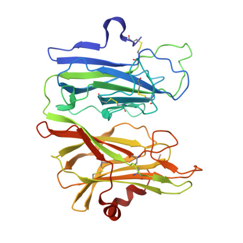
Many bioactive peptides, such as hormones and neuropeptides, require amidation at the C terminus for their full biological activity. Peptidylglycine α-hydroxylating monooxygenase (PHM) performs the first step of the amidation reaction-the hydroxylation of peptidylglycine substrates at the Cα position of the terminal glycine. The hydroxylation reaction is copper- and O(2)-dependent and requires 2 equiv of exogenous reductant. The proposed mechanism suggests that O(2) is reduced by two electrons, each provided by one of two nonequivalent copper sites in PHM (Cu(H) and Cu(M)). The characteristics of the reduced oxygen species in the PHM reaction and the identity of the reactive intermediate remain uncertain. To further investigate the nature of the key intermediates in the PHM cycle, we determined the structure of the oxidized form of PHM complexed with hydrogen peroxide. In this 1.98-Å-resolution structure (hydro)peroxide binds solely to Cu(M) in a slightly asymmetric side-on mode. The O-O interatomic distance of the copper-bound ligand is 1.5 Å, characteristic of peroxide/hydroperoxide species, and the Cu-O distances are 2.0 and 2.1 Å. Density functional theory calculations using the first coordination sphere of the Cu(M) active site as a model system show that the computed energies of the side-on L(3)Cu(M)(II)-O(2) (2-) species and its isomeric, end-on structure L(3)Cu(M)(I)-O(2) (·-) are similar, suggesting that both these intermediates are significantly populated within the protein environment. This observation has important mechanistic implications. The geometry of the observed side-on coordinated peroxide ligand in L(3)Cu(M)(II)O(2) (2-) is in good agreement with the results of a hybrid quantum mechanical-molecular mechanical optimization of this species.
Organizational Affiliation:
Department of Biophysics and Biophysical Chemistry, Johns Hopkins School of Medicine, Johns Hopkins University, Baltimore, MD, 21205, USA.