4DSH

Crystal structure of reduced UDP-Galactopyranose mutase


Experimental Data Snapshot

  • Method: X-RAY DIFFRACTION
  • Resolution: 2.25 Å
  • R-Value Free: 0.210 
  • R-Value Work: 0.180 
  • R-Value Observed: 0.182 

wwPDB Validation   3D Report Full Report


Ligand Structure Quality Assessment 


This is version 1.2 of the entry. See complete history


Literature

Crystal Structures of Trypanosoma cruzi UDP-Galactopyranose Mutase Implicate Flexibility of the Histidine Loop in Enzyme Activation.

Dhatwalia, R.Singh, H.Oppenheimer, M.Sobrado, P.Tanner, J.J.

(2012) Biochemistry 51: 4968-4979

  • DOI: https://doi.org/10.1021/bi300498c
  • Primary Citation of Related Structures:  
    4DSG, 4DSH

  • PubMed Abstract: 

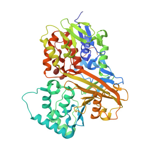
    Chagas disease is a neglected tropical disease caused by the protozoan parasite Trypanosoma cruzi. Here we report crystal structures of the galactofuranose biosynthetic enzyme UDP-galactopyranose mutase (UGM) from T. cruzi, which are the first structures of this enzyme from a protozoan parasite. UGM is an attractive target for drug design because galactofuranose is absent in humans but is an essential component of key glycoproteins and glycolipids in trypanosomatids. Analysis of the enzyme-UDP noncovalent interactions and sequence alignments suggests that substrate recognition is exquisitely conserved among eukaryotic UGMs and distinct from that of bacterial UGMs. This observation has implications for inhibitor design. Activation of the enzyme via reduction of the FAD induces profound conformational changes, including a 2.3 Å movement of the histidine loop (Gly60-Gly61-His62), rotation and protonation of the imidazole of His62, and cooperative movement of residues located on the si face of the FAD. Interestingly, these changes are substantially different from those described for Aspergillus fumigatus UGM, which is 45% identical to T. cruzi UGM. The importance of Gly61 and His62 for enzymatic activity was studied with the site-directed mutant enzymes G61A, G61P, and H62A. These mutations lower the catalytic efficiency by factors of 10-50, primarily by decreasing k(cat). Considered together, the structural, kinetic, and sequence data suggest that the middle Gly of the histidine loop imparts flexibility that is essential for activation of eukaryotic UGMs. Our results provide new information about UGM biochemistry and suggest a unified strategy for designing inhibitors of UGMs from the eukaryotic pathogens.


  • Organizational Affiliation

    Department of Chemistry, University of Missouri-Columbia, Columbia, Missouri 65211, USA.


Macromolecules
Find similar proteins by:  (by identity cutoff)  |  3D Structure
Entity ID: 1
MoleculeChains Sequence LengthOrganismDetailsImage
UDP-galactopyranose mutase
A, B
484Trypanosoma cruziMutation(s): 0 
Gene Names: Tc00.1047053507993.160
EC: 5.4.99.9
UniProt
Find proteins for Q4E1W2 (Trypanosoma cruzi (strain CL Brener))
Explore Q4E1W2 
Go to UniProtKB:  Q4E1W2
Entity Groups  
Sequence Clusters30% Identity50% Identity70% Identity90% Identity95% Identity100% Identity
UniProt GroupQ4E1W2
Sequence Annotations
Expand
  • Reference Sequence
Small Molecules
Ligands 4 Unique
IDChains Name / Formula / InChI Key2D Diagram3D Interactions
FDA
Query on FDA

Download Ideal Coordinates CCD File 
D [auth A],
I [auth B]
DIHYDROFLAVINE-ADENINE DINUCLEOTIDE
C27 H35 N9 O15 P2
YPZRHBJKEMOYQH-UYBVJOGSSA-N
UDP
Query on UDP

Download Ideal Coordinates CCD File 
C [auth A],
J [auth B]
URIDINE-5'-DIPHOSPHATE
C9 H14 N2 O12 P2
XCCTYIAWTASOJW-XVFCMESISA-N
EPE
Query on EPE

Download Ideal Coordinates CCD File 
H [auth A]4-(2-HYDROXYETHYL)-1-PIPERAZINE ETHANESULFONIC ACID
C8 H18 N2 O4 S
JKMHFZQWWAIEOD-UHFFFAOYSA-N
SO4
Query on SO4

Download Ideal Coordinates CCD File 
E [auth A],
F [auth A],
G [auth A],
K [auth B]
SULFATE ION
O4 S
QAOWNCQODCNURD-UHFFFAOYSA-L
Experimental Data & Validation

Experimental Data

  • Method: X-RAY DIFFRACTION
  • Resolution: 2.25 Å
  • R-Value Free: 0.210 
  • R-Value Work: 0.180 
  • R-Value Observed: 0.182 
  • Space Group: P 65 2 2
Unit Cell:
Length ( Å )Angle ( ˚ )
a = 143.84α = 90
b = 143.84β = 90
c = 354.389γ = 120
Software Package:
Software NamePurpose
XSCALEdata scaling
PHENIXrefinement
PDB_EXTRACTdata extraction
ADSCdata collection
XDSdata reduction
MOLREPphasing

Structure Validation

View Full Validation Report


Ligand Structure Quality Assessment 


Entry History 

Deposition Data

Revision History  (Full details and data files)

  • Version 1.0: 2012-06-13
    Type: Initial release
  • Version 1.1: 2012-09-05
    Changes: Database references
  • Version 1.2: 2023-09-13
    Changes: Data collection, Database references, Derived calculations, Refinement description