4DQH

Crystal Structure of (R14C/E65C) HIV-1 Protease in complex with DRV


Experimental Data Snapshot

  • Method: X-RAY DIFFRACTION
  • Resolution: 1.79 Å
  • R-Value Free: 0.219 
  • R-Value Work: 0.170 
  • R-Value Observed: 0.173 

wwPDB Validation   3D Report Full Report


Ligand Structure Quality Assessment 


This is version 1.3 of the entry. See complete history


Literature

Hydrophobic core flexibility modulates enzyme activity in HIV-1 protease.

Mittal, S.Cai, Y.Nalam, M.N.Bolon, D.N.Schiffer, C.A.

(2012) J Am Chem Soc 134: 4163-4168

  • DOI: https://doi.org/10.1021/ja2095766
  • Primary Citation of Related Structures:  
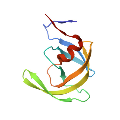
    4DQB, 4DQC, 4DQE, 4DQF, 4DQG, 4DQH

  • PubMed Abstract: 

    Human immunodeficiency virus Type-1 (HIV-1) protease is crucial for viral maturation and infectivity. Studies of protease dynamics suggest that the rearrangement of the hydrophobic core is essential for enzyme activity. Many mutations in the hydrophobic core are also associated with drug resistance and may modulate the core flexibility. To test the role of flexibility in protease activity, pairs of cysteines were introduced at the interfaces of flexible regions remote from the active site. Disulfide bond formation was confirmed by crystal structures and by alkylation of free cysteines and mass spectrometry. Oxidized and reduced crystal structures of these variants show the overall structure of the protease is retained. However, cross-linking the cysteines led to drastic loss in enzyme activity, which was regained upon reducing the disulfide cross-links. Molecular dynamics simulations showed that altered dynamics propagated throughout the enzyme from the engineered disulfide. Thus, altered flexibility within the hydrophobic core can modulate HIV-1 protease activity, supporting the hypothesis that drug resistant mutations distal from the active site can alter the balance between substrate turnover and inhibitor binding by modulating enzyme activity.


  • Organizational Affiliation

    Department of Biochemistry and Molecular Pharmacology, University of Massachusetts Medical School, Worcester, Massachusetts 01605, USA.


Macromolecules
Find similar proteins by:  (by identity cutoff)  |  3D Structure
Entity ID: 1
MoleculeChains Sequence LengthOrganismDetailsImage
Wild-type HIV-1 protease dimer
A, B
99Human immunodeficiency virus 1Mutation(s): 0 
Gene Names: gag-pol
UniProt
Find proteins for P12499 (Human immunodeficiency virus type 1 group M subtype D (isolate Z2/CDC-Z34))
Explore P12499 
Go to UniProtKB:  P12499
Entity Groups  
Sequence Clusters30% Identity50% Identity70% Identity90% Identity95% Identity100% Identity
UniProt GroupP12499
Sequence Annotations
Expand
  • Reference Sequence
Small Molecules
Ligands 2 Unique
IDChains Name / Formula / InChI Key2D Diagram3D Interactions
017
Query on 017

Download Ideal Coordinates CCD File 
C [auth B](3R,3AS,6AR)-HEXAHYDROFURO[2,3-B]FURAN-3-YL(1S,2R)-3-[[(4-AMINOPHENYL)SULFONYL](ISOBUTYL)AMINO]-1-BENZYL-2-HYDROXYPROPYLCARBAMATE
C27 H37 N3 O7 S
CJBJHOAVZSMMDJ-HEXNFIEUSA-N
GOL
Query on GOL

Download Ideal Coordinates CCD File 
D [auth B]GLYCEROL
C3 H8 O3
PEDCQBHIVMGVHV-UHFFFAOYSA-N
Binding Affinity Annotations 
IDSourceBinding Affinity
017 BindingDB:  4DQH Ki: min: 2.00e-4, max: 2 (nM) from 17 assay(s)
Kd: 0.02 (nM) from 1 assay(s)
IC50: min: 0.06, max: 370 (nM) from 10 assay(s)
EC50: min: 0.15, max: 112 (nM) from 5 assay(s)
Experimental Data & Validation

Experimental Data

  • Method: X-RAY DIFFRACTION
  • Resolution: 1.79 Å
  • R-Value Free: 0.219 
  • R-Value Work: 0.170 
  • R-Value Observed: 0.173 
  • Space Group: P 21 21 21
Unit Cell:
Length ( Å )Angle ( ˚ )
a = 50.706α = 90
b = 57.982β = 90
c = 61.546γ = 90
Software Package:
Software NamePurpose
DENZOdata reduction
SCALEPACKdata scaling
REFMACrefinement
PDB_EXTRACTdata extraction

Structure Validation

View Full Validation Report


Ligand Structure Quality Assessment 


Entry History 

Deposition Data

Revision History  (Full details and data files)

  • Version 1.0: 2012-03-07
    Type: Initial release
  • Version 1.1: 2012-05-02
    Changes: Database references
  • Version 1.2: 2017-11-15
    Changes: Refinement description
  • Version 1.3: 2024-02-28
    Changes: Data collection, Database references, Derived calculations, Structure summary