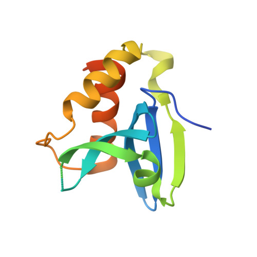
Structural Basis for the Acyltransferase Activity of Lecithin:Retinol Acyltransferase-like Proteins.
Golczak, M., Kiser, P.D., Sears, A.E., Lodowski, D.T., Blaner, W.S., Palczewski, K.(2012) J Biol Chem 287: 23790-23807
- PubMed: 22605381
- DOI: https://doi.org/10.1074/jbc.M112.361550
- Primary Citation of Related Structures:
4DOT, 4DPZ - PubMed Abstract:
Lecithin:retinol acyltransferase-like proteins, also referred to as HRAS-like tumor suppressors, comprise a vertebrate subfamily of papain-like or NlpC/P60 thiol proteases that function as phospholipid-metabolizing enzymes. HRAS-like tumor suppressor 3, a representative member of this group, plays a key role in regulating triglyceride accumulation and energy expenditure in adipocytes and therefore constitutes a novel pharmacological target for treatment of metabolic disorders causing obesity. Here, we delineate a catalytic mechanism common to lecithin:retinol acyltransferase-like proteins and provide evidence for their alternative robust lipid-dependent acyltransferase enzymatic activity. We also determined high resolution crystal structures of HRAS-like tumor suppressor 2 and 3 to gain insight into their active site architecture. Based on this structural analysis, two conformational states of the catalytic Cys-113 were identified that differ in reactivity and thus could define the catalytic properties of these two proteins. Finally, these structures provide a model for the topology of these enzymes and allow identification of the protein-lipid bilayer interface. This study contributes to the enzymatic and structural understanding of HRAS-like tumor suppressor enzymes.
Organizational Affiliation:
Department of Pharmacology, School of Medicine, Case Western Reserve University, Cleveland, Ohio 44106, USA. mxg149@case.edu